. 2010 Dec 9;468(7325):784-9.
doi: 10.1038/nature09516. Epub 2010 Nov 14.

Structure of a bacterial ribonuclease P holoenzyme in complex with tRNA

Affiliations

Structure of a bacterial ribonuclease P holoenzyme in complex with tRNA

Nicholas J Reiter et al. Nature. .

Abstract

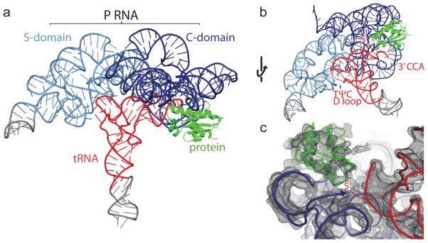
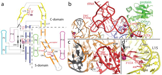
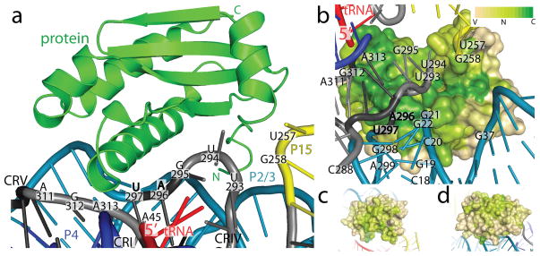
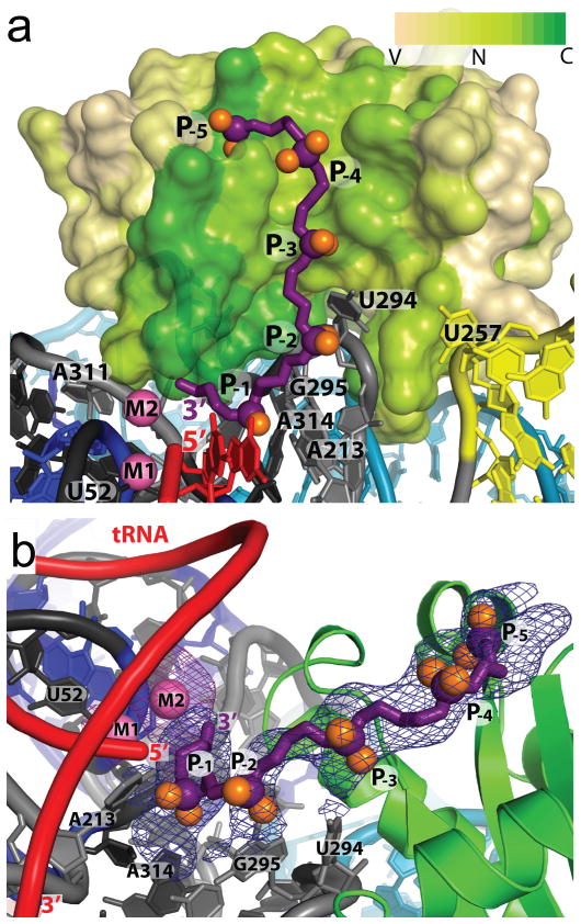
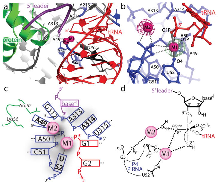
Ribonuclease (RNase) P is the universal ribozyme responsible for 5'-end tRNA processing. We report the crystal structure of the Thermotoga maritima RNase P holoenzyme in complex with tRNA(Phe). The 154 kDa complex consists of a large catalytic RNA (P RNA), a small protein cofactor and a mature tRNA. The structure shows that RNA-RNA recognition occurs through shape complementarity, specific intermolecular contacts and base-pairing interactions. Soaks with a pre-tRNA 5' leader sequence with and without metal help to identify the 5' substrate path and potential catalytic metal ions. The protein binds on top of a universally conserved structural module in P RNA and interacts with the leader, but not with the mature tRNA. The active site is composed of phosphate backbone moieties, a universally conserved uridine nucleobase, and at least two catalytically important metal ions. The active site structure and conserved RNase P-tRNA contacts suggest a universal mechanism of catalysis by RNase P.


Conflict of interest statement

Competing Interests statement The authors declare that they have no competing financial interests.

Figures

Figure 1
Figure 1. Crystal structure of the T. maritima RNase P holoenzyme in complex with tRNA
a, Structure of bacterial RNase P, composed of a large RNA subunit (338 nucleotides, ~110 kDa) and a small protein component (117 amino acids, ~14.3 kDa), in complex with tRNA (76 nucleotides, ~26 kDa). The RNA component serves as the primary biocatalyst in the reaction and contains two domains, termed the catalytic (C, blue) and specificity (S, light blue) domains. The RNase P protein (green) binds the 5′ leader region of the pre-tRNA substrate and assists in product release. Transfer RNA (tRNAPhe) (red) makes multiple interactions with the P RNA (see Fig. 2 and S1 for details). Regions in grey denote additional RNA nucleotides required for crystallization. b, Alternate view of the RNase P/tRNA complex, identifying the tRNA recognition regions: the 5′ end where catalysis occurs, the 3′ CCA end, and the highly conserved TΨC and D loop regions. c, View of the 4.1 Å experimental electron density map centered on the 5′ end of tRNA. The map is represented as a dark grey mesh, contoured at 1.4 rmsd.
Figure 2
Figure 2. tRNA recognition by RNase P is mediated by RNA-RNA interactions
a, Schematic of the P RNA secondary structure mapping the tRNA-P RNA contacts observed in the crystal structure. The tRNA nucleotides (1•72, 2, 3, 64, and 65) and regions (5′, 3′, TΨC loop, D loop, and acceptor) involved in direct interactions are shown in red. Intermolecular base pairs form between the 3′ end of tRNA (DCCA) and loop 15 (L15), where D is the discriminator nucleotide that serves as an identity element in tRNA biogenesis. P RNA nucleotides that are universally conserved (black, uppercase), conserved among all bacteria (grey, uppercase), or highly conserved in bacteria (black, lowercase) are identified. Metal ions are shown as filled pink circles, and denote the location of the active site (M1, M2), and other structurally important regions (M3, M4). Single and double dashes in red represent minor groove and base stacking interactions, respectively. All identified tRNA/P RNA contacts are within 4 Å. The crystallized T. maritima P RNA consists of eighteen paired helices (P), five universally conserved regions (CRI to CR-V) (black), two junctions containing conserved nucleotides in bacteria (dark grey), several loop (L) regions, and an engineered tetraloop region (T, light grey). The coaxial P1/P4/P5 stem is shown in blue, P2/P3 stems in cyan, P6/P15/P16 and L15/L17 in yellow, P7 and P10/P11/P12 in orange, P8/P9 in light green, and P13/P14 in pink (see Fig. S1 for additional details). b, Recognition of tRNA by the P RNA of RNase P. The acceptor stem of tRNA (red) docks onto the P RNA (colored as in a) making a series of interactions, including base stacking in the TΨC/D loops of tRNA and the S-domain, an A-minor interaction, and base pairing, ribose zippers, and stacking interactions between the 5′ and 3′ ends of tRNA and the C-domain. The protein (green) makes no direct contacts with mature tRNA. Critical metals ions (M1–M4) identified are shown as magenta spheres. c, tRNA recognition by the S-domain. Two universally conserved P RNA regions (CR-II and III, dark grey) facilitate base stacking interactions with unstacked bases in the structurally conserved TΨC and D loops of tRNA. Dashed circles highlight this stacking interaction between P RNA residues A112, G147 and tRNA residues G19, C56. A conserved P RNA adenosine (A198) stacks into the minor groove of the acceptor tRNA stem. d, Recognition of the tRNA 3′ CCA by the C-domain. Intermolecular base pairs form between the 3′ tRNA (ACC) and the L15 (GGU) loop of P RNA. This interaction is stabilized by a structural metal (M3, magenta sphere) and a L15 ribose zipper conformation.
Figure 3
Figure 3. Protein-RNA contacts within the RNase P holoenzyme
a, The protein sits on the P RNA surface formed by conserved regions I, IV, and V. The protein (green, shown as ribbons) additionally contacts the L15/P15 junction and the P2/3 helices (P RNA as colored in Fig. 2). Labeled P RNA nucleotides make protein contacts (within 4 Å) and include: A45 in CR-I, U257 and G258 in the L15/P15 junction, U293, U294, G295, A296, U297 in CR-IV, and A311, G312, and A313 in CR-V. Bold nucleotides are universally conserved. b, Surface representation of the protein colored by sequence conservation (Variable: tan, Neutral: light green, Conserved: green). A highly conserved patch in the protein extends from the vicinity of the 5′ end of the tRNA, and interacts with P RNA conserved regions IV (U293–U297) and V (A311–A313). Other P RNA nucleotides that make protein contacts include: the P2 helix (C18-G22, G298-A299), the P3 helix (G37), and the L15/P15 junction (U257-G258). Four hundred and ninety bacterial RNase P proteins were included in the analysis of the sequence conservation using the ConSurf server. Panels (c) and (d) show different orientations to emphasize that high sequence conservation is concentrated in the region of the protein that faces the conserved regions of the P RNA. Neutral or slightly conserved regions shown in these two orientations correspond to a patch that interacts with the leader.
Figure 4
Figure 4. Pre-tRNA leader - protein interactions in the RNase P holoenzyme
a, Surface representation of the protein colored by sequence conservation as in Fig. 3. The pre-tRNA 5′ leader (purple, with purple and orange spheres for the phosphorous and non-bridging oxygens, respectively) was modeled as a polyphosphate chain with five phosphates (P-1 to P-5). The leader follows a highly conserved patch in the protein extending from the 5′ end of the mature tRNA (red) and away from the P RNA. The addition of a 5′ leader with metal (Sm3+) reveals a second metal ion (M2). b, Alternative view of the pre-tRNA leader/protein interaction. Each phosphate position (P-1 though P-5) was visible in a 4.2 Å difference Fourier map (mFo-DFc) calculated from crystals where only the leader was soaked into the crystals (blue mesh, 3 rmsd contour levels). A second 4.2 Å difference Fourier map (mFo-DFc) calculated from crystals where the leader and Sm3+ metal were soaked into the crystals shows clearly the position of the second metal ion (magenta mesh, 3.5 rmsd contour level). P RNA residues poised to make contacts are labeled. Nucleotide U52 serves as a reference point in a and b and does not interact with the 5′ leader oligonucleotide.
Figure 5
Figure 5. Structure of the RNase P active site environment
a, The active site is inferred from the location of the mature 5′ end of tRNA. The diagram shows the position of the mature tRNA (red), the leader (purple), the protein component (green), and the P RNA (blue and grey). A group of conserved P RNA nucleotides (A49 - U52, A213, A313, and A314) form part of the active site. Two metal ions (magenta spheres) are found in the active site. b, The two active site metal ions (M1 and M2) are within 4 Å of the 5′ phosphate of tRNA and the M1–M2 metal-metal distance is ~4.8 Å. The M1 metal makes contacts (≤2.1 Å, solid grey bonds, labeled) with tRNA (G1 O1P) and P RNA (A50 O1P and U52 O4) oxygens. Other possible ligands within 3.5 Å of M1 or M2 are represented by dashed grey lines (Table STV). The figure shows two isomorphous difference Fourier (mFo-DFc) maps. The green mesh corresponds to a Eu3+ soak in the absence of leader and is contoured at the 9.5 rmsd level. The magenta mesh corresponds to a Sm3+ and 5′ leader soak and is contoured at the 5.5 rmsd level. The second metal is clearly visible only when the leader is present. c, Schematic diagram of the interactions around the active site. The diagram shows all residues within 8 Å of the 5′ phosphorus atom of tRNA. Short dashed lines represent metal ligand distances within 2.2 Å and longer dashed lines represent nucleotides which form canonical base pairs. Nucleotides in bold are universally conserved in P RNA. The P RNA, tRNA, 5′ leader, and protein side chains are shown in blue, red, purple and green, respectively. d, Proposed reaction mechanism for the endonucleoytic cleavage of pre-tRNA by RNase P based on the structure of the enzyme-product (E-P) complex and previous mechanistic studies,. The M1 metal distance to the 5′ phosphate ligands (Table STV) in the E-P complex is consistent with the proposed enzyme-substrate (E-S) transition state. In this proposed reaction scheme, M1 is ~180° from the apical O3′ position and activates a hydroxyl nucleophile for an in-line nucleophillic displacement, creating a new bond and displacing the 3′ scissile phosphate oxygen. As RNase P proceeds through an SN2 reaction pathway, the stereochemistry around the phosphorus atom undergoes a net inversion of configuration. If the pro-RP (O2P) oxygen coordinates metal in the E-S complex during catalysis, as previously observed,, this would subsequently allow for the pro-SP (O1P) oxygen to coordinate metal in the E-P complex, as observed in the crystal structure. Product release could be facilitated by a metal (M2) coordinated water, which would enable proton transfer to the 3′ scissile oxygen. The exact active site geometry and identity of other metal ligands in an E-S complex has yet to be established.

Comment in

  • The RNP bridge between two worlds.
    Schimmel P. Schimmel P. Nat Rev Mol Cell Biol. 2011 Mar;12(3):135. doi: 10.1038/nrm3061. Epub 2011 Feb 2. Nat Rev Mol Cell Biol. 2011. PMID: 21285979 No abstract available.

References

    1. Hartmann RK, Gossringer M, Spath B, Fischer S, Marchfelder A. The making of tRNAs and more - RNase P and tRNase Z. Prog Mol Biol Transl Sci. 2009;85:319–368. - PubMed
    1. Kazantsev AV, Pace NR. Bacterial RNase P: a new view of an ancient enzyme. Nat Rev Microbiol. 2006;4:729–740. - PubMed
    1. Liu F, Altman S. Ribonuclease P. Vol. 10. Springer Science+Business Media, LLC; 2010.
    1. Guerrier-Takada C, Gardiner K, Marsh T, Pace N, Altman S. The RNA moiety of ribonuclease P is the catalytic subunit of the enzyme. Cell. 1983;35:849–857. - PubMed
    1. Pan T, Loria A, Zhong K. Probing of tertiary interactions in RNA: 2′-hydroxyl-base contacts between the RNase P RNA and pre-tRNA. Proc Natl Acad Sci USA. 1995;92:12510–12514. - PMC - PubMed

Publication types

MeSH terms