

doi: 10.1038/nmeth.1533.
Epub 2010 Nov 14.
Three-dimensional cellular ultrastructure resolved by X-ray microscopy
Affiliations
- PMID: 21076419
- PMCID: PMC7337972
- DOI: 10.1038/nmeth.1533
Item in Clipboard
Three-dimensional cellular ultrastructure resolved by X-ray microscopy
Nat Methods.
2010 Dec.
Abstract
We developed an X-ray microscope using partially coherent object illumination instead of previously used quasi-incoherent illumination. The design permitted the incorporation of a cryogenic tilt stage, enabling tomography of frozen-hydrated, intact adherent cells. We obtained three-dimensional reconstructions of mouse adenocarcinoma cells at ∼36-nm (Rayleigh) and ∼70-nm (Fourier ring correlation) resolution, which allowed us to visualize the double nuclear membrane, nuclear pores, nuclear membrane channels, mitochondrial cristae and lysosomal inclusions.
Conflict of interest statement
COMPETING FINANCIAL INTERESTS
The authors declare no competing financial interests.
Figures
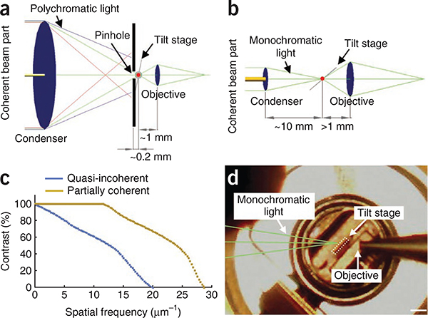
X-ray microscope designs. (a) Schematic of the design for quasi-incoherent imaging, in which the X-ray source produces a divergent X-ray beam containing small coherently illuminated areas (dim yellow), which are a tiny fraction of the total beam and condenser diameter (~10 mm). The numerical apertures (NA) of the condenser and the objective (both of which are zone plates) were matched (NAcondenser / NAobjective − 1), and a 50-nm zone plate objective is used. (b) Schematic of the design for partially coherent imaging, in which the X-ray source produces a more collimated photon beam containing ~100× more coherent light (bright yellow) with a coherent beam diameter that is a substantial fraction of the condenser diameter (2 mm). The numerical aperture of the objective is more than twice that of the condenser (NAcondenser / NAobjective − 0.43). A 25-nm zone plate objective is used. (c) Contrast transfer functions are plotted for partially coherent and quasi-incoherent microscope designs. The curves were calculated theoretically using the optical parameters of the two designs. (d) Photograph of the tilted flat sample holder (tilt stage) inside the microscope chamber. Scale bar, 1 mm.
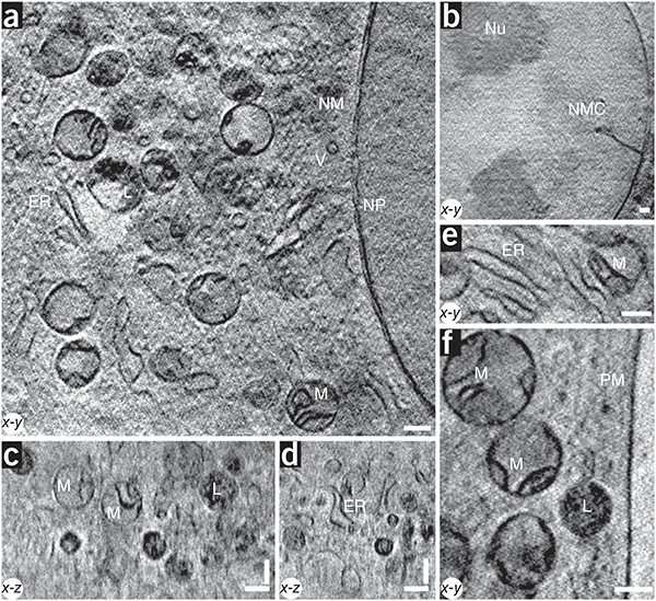
X-ray images of a cell. (a–f) The 3D partially coherent X-ray tomograms of mouse adenocarcinoma cells show many subcellular organelles including mitochondria (M), lyosomes (L), endoplasmic reticulum (ER), vesicles (V), the plasma membrane (PM), the nuclear membrane (NM), nuclear pores (NP), nucleoli (Nu) and nuclear membrane channels (NMC). All images were acquired with a 25-nm zone plate at 510 eV photon energy, except for the image shown in b, which was acquired with a 40-nm zone plate. Pixel sizes and slice thicknesses are 9.8 nm (a,c–f) and 15.6 nm (b). Scale bars, 0.39 μm.
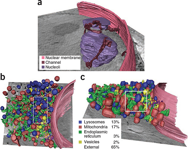
Volumetric rendering of cell cytoplasm. (a) The 3D data corresponding to the image in Figure 2b was segmented to visualize the association of the nuclear membrane channels with the nuclear membrane. (b,c) The 3D data corresponding to the image in Figure 2a were segmented, yielding x-y (b) and x-z (c) views of the cytoplasm. Percentages indicate the volume fraction occupied by different organelles measured in the 3D subvolume delineated by the white rectangles.
References
-
- Schneider G et al. Surf. Rev. Lett. 9, 177–183 (2002).
Publication types
MeSH terms
Grants and funding
LinkOut - more resources
Full Text Sources
Other Literature Sources
