. 2011:2011:803015.
doi: 10.1155/2011/803015. Epub 2010 Nov 4.

Neuroprotection by the Traditional Chinese Medicine, Tao-Hong-Si-Wu-Tang, against Middle Cerebral Artery Occlusion-Induced Cerebral Ischemia in Rats

Affiliations

Neuroprotection by the Traditional Chinese Medicine, Tao-Hong-Si-Wu-Tang, against Middle Cerebral Artery Occlusion-Induced Cerebral Ischemia in Rats

Chih-Jen Wu et al. Evid Based Complement Alternat Med. 2011.

Abstract

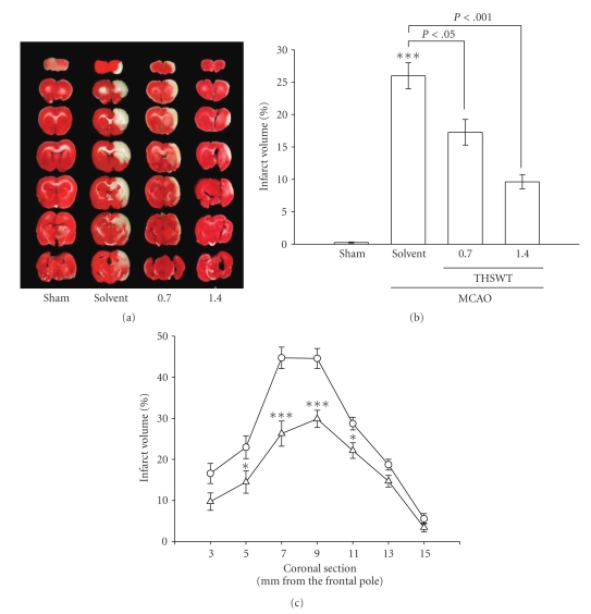
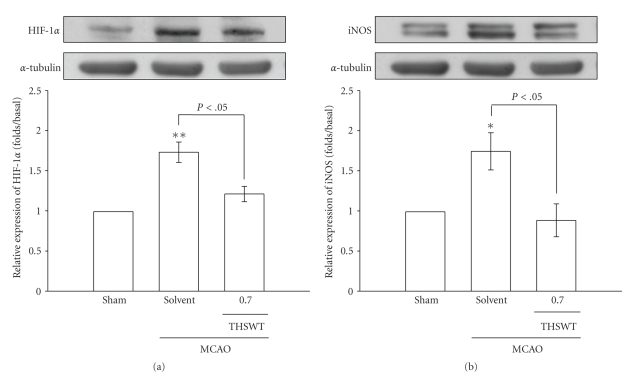
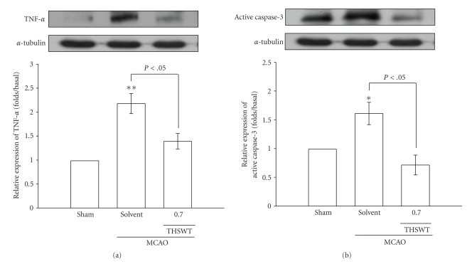
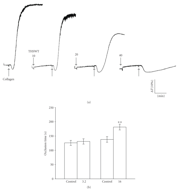
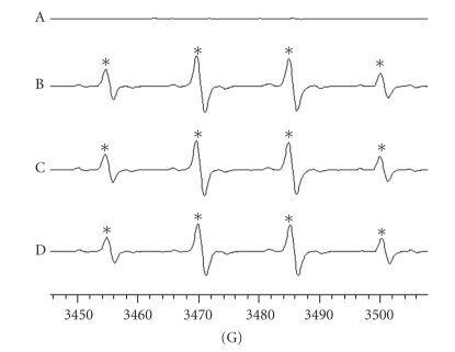
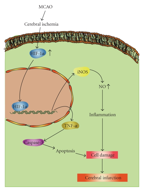
Tao-Hong-Si-Wu-Tang (THSWT) is a famous traditional Chinese medicine (TMC). In the present study, oral administration of THSWT (0.7 and 1.4 g kg(-1)day(-1)) for 14 days before MCAO dose-dependently attenuated focal cerebral ischemia in rats. MCAO-induced focal cerebral ischemia was associated with increases in hypoxia-inducible factor (HIF)-1α, inducible nitric oxide synthase (iNOS), tumor necrosis factor (TNF)-α, and active caspase-3 expressions in ischemic regions. These expressions were obviously inhibited by 0.7 g kg(-1)day(-1) THSWT treatment. In addition, THSWT inhibited platelet aggregation stimulated by collagen in washed platelets. In an in vivo study, THSWT (16 g kg(-1)) significantly prolonged platelet plug formation in mice. However, THSWT (20 and 40 μg mL(-1)) did not significantly reduce the electron spin resonance (ESR) signal intensity of hydroxyl radical (OH(•)) formation. In conclusion, the most important findings of this study demonstrate for the first time that THSWT possesses potent neuroprotective activity against MCAO-induced focal cerebral ischemia in vivo. This effect may be mediated, at least in part, by the inhibition of both HIF-1α and TNF-α activation, followed by the inhibition of inflammatory responses (i.e., iNOS expression), apoptosis formation (active caspase-3), and platelet activation, resulting in a reduction in the infarct volume in ischemia-reperfusion brain injury.


Figures

Figure 1
Figure 1
Effects of Tao-Hong-Si-Wu-Tang (THSWT) in middle cerebral artery occlusion-(MCAO-) induced focal cerebral ischemia in rats. (a) Coronal sections of 2,3,5-triphenyltetrazolium-(TTC-) stained brains, (b) dose-response effect of THSWT, and (c) the infarct area at various distances from the frontal pole 24 h after MACO-reperfusion rats. Cerebral infarction in sham-operated (sham) or MACO-reperfusion rats is from a representative animal that received the solvent (distilled water) or THSWT (0.7 and 1.4 g kg−1day−1) orally for 14 days. (a) TTC-stained brains and (b) infarct volumes were calculated as described in “Methods”, and data are presented as the infarct volume for each animal in the group as well as the means ± S.E.M. (n = 7). ***P < .001, compared to the sham-operated group. (c) Forebrain profiles of the infarct area at various distances from the frontal pole as described in “Methods”. Each point (○, solvent-treated group; Δ, THSWT 0.7 g kg−1day−1-treated group) and vertical bar represent the means ± S.E.M. (n = 7). *P < .05 and ***P < .001, compared to the solvent-treated group.
Figure 2
Figure 2
Effects of Tao-Hong-Si-Wu-Tang (THSWT) on the expressions of (a) hypoxia-inducible factor (HIF-1α and (b) inducible nitric oxide synthase (iNOS) in cerebral homogenates 24 h after middle cerebral artery occlusion-(MCAO-) reperfusion injury in rats. Fresh brains from sham-operated (lane 1), solvent-treated (lane 2), and THSWT- (0.7 g kg−1day−1) treated (lane 3) rats were removed and sectioned coronally into four sequential parts from the frontal lobe to the occipital lobe. The third of four sequential parts of the ischemic-injured hemisphere was separately collected, homogenized, and centrifuged. The supernatant (50 μg protein) was then subjected to SDS-PAGE, and transferred onto membranes for analysis of HIF-1α and iNOS expressions. The results are representative examples of three similar experiments. Data are presented as the means ± S.E.M. *P < .05 and **P < .01, compared to the sham-operated group (lane 1). Equal loading in each lane is demonstrated by similar intensities of α-tubulin.
Figure 3
Figure 3
Effects of Tao-Hong-Si-Wu-Tang (THSWT) on the expressions of (a) tumor necrosis factor (TNF)-α and (b) active caspase-3 in cerebral homogenates 24 h after middle cerebral artery occlusion-(MCAO-) reperfusion injury in rats. Fresh brains from sham-operated (lane 1), solvent-treated (lane 2), and THSWT (0.7 g kg−1day−1)-treated (lane 3) rats were removed and sectioned coronally into four sequential parts from the frontal lobe to the occipital lobe. The third of four sequential parts of the ischemic-injured hemisphere was separately collected, homogenized, and centrifuged. The supernatant (50 μg protein) was then subjected to SDS-PAGE and transferred onto membranes for analysis of TNF-α and active caspase-3 expressions. The results are representative examples of three similar experiments. Data are presented as the means ± S.E.M. *P < .05 and **P < .01, compared to the sham-operated group (lane 1). Equal loading in each lane is demonstrated by similar intensities of α-tubulin.
Figure 4
Figure 4
Inhibitory effect of Tao-Hong-Si-Wu-Tang (THSWT) on platelet aggregation in vitro and prolongation of the occlusion time for inducing thrombus formation in vivo. (a) Washed human platelets (3.6 × 10⁸ platelets mL−1) were preincubated with THSWT (20 and 40 μg ml−1) for 3 min, followed by the addition of collagen (1 μg mL−1) to trigger platelet aggregation. The profiles are a representative example of three similar experiments. (b) Mice were administered the solvent control (normal saline) or THSWT (3.2 and 16 g kg−1), after which mesenteric venules were selected for irradiation to induce microthrombus formation. Data of the bar graphs in (b) are presented as the means ± S.E.M. of the occlusion time (s) for inducing platelet plug formation (n = 4). **P < .01, compared to the individual solvent control group.
Figure 5
Figure 5
Inhibition of the free radical-scavenging activity by Tao-Hong-Si-Wu-Tang (THSWT) in the H2O2/NaOH/DMSO system. The signal of hydroxyl radical peaks was observed in electron spin resonance (ESR) experiments. (a) Resting spectrum (without H2O2), (b) typical ESR spectra in the presence of solvent control (distilled water), (c) THSWT (20 μg mL−1), and (d) (40 μg ml−1) in the H2O2/NaOH/DMSO system. The spectrum is a representative example of four similar experiments. An asterisk (*) indicates formation of hydroxyl radicals.
Figure 6
Figure 6
Hypothetical scheme of middle cerebral artery occlusion-(MCAO-) induced focal cerebral ischemia. MCAO decreases the blood flow of ischemic area followed by inducing a release of transcription factor HIF-1α . HIF-1α translocates into the nucleus in activating genes of iNOS and TNF-α. iNOS may contribute to enhance the inflammatory responses by elevating nitric oxide (NO). TNF-α, it shows to be involved in apoptosis through the activation of downstream caspases-3, and subsequently induces cerebral infarction.

References

    1. Kuroda S, Siesjö BK. Reperfusion damage following focal ischemia: pathophysiology and therapeutic windows. Clinical Neuroscience. 1997;4(4):199–212. - PubMed
    1. Abumiya T, Fitridge R, Mazur C, et al. Integrin α(IIb)β3 inhibitor preserves microvascular patency in experimental acute focal cerebral ischemia. Stroke. 2000;31(6):1402–1410. - PubMed
    1. Iadecola C, Zhang F, Casey R, Clark HB, Ross ME. Inducible nitric oxide synthase gene expression in vascular cells after transient focal cerebral ischemia. Stroke. 1996;27(8):1373–1380. - PubMed
    1. Chan PH. Reactive oxygen radicals in signaling and damage in the ischemic brain. Journal of Cerebral Blood Flow and Metabolism. 2001;21(1):2–14. - PubMed
    1. Lipton P. Ischemic cell death in brain neurons. Physiological Reviews. 1999;79(4):1431–1568. - PubMed