

Chest radiographic findings in primary pulmonary tuberculosis: observations from high school outbreaks
- PMID: 21076586
- PMCID: PMC2974222
- DOI: 10.3348/kjr.2010.11.6.612
Chest radiographic findings in primary pulmonary tuberculosis: observations from high school outbreaks
Abstract
Objective: To describe the radiographic findings of primary pulmonary tuberculosis (TB) in previously healthy adolescent patients.
Materials and methods: The Institutional Review Board approved this retrospective study, with a waiver of informed consent from the patients. TB outbreaks occurred in 15 senior high schools and chest radiographs from 58 students with identical strains of TB were analyzed by restriction fragment length polymorphism analysis by two independent observers. Lesions of nodule(s), consolidation, or cavitation in the upper lung zones were classified as typical TB. Mediastinal lymph node enlargement; lesions of nodule(s), consolidation, or cavitation in lower lung zones; or pleural effusion were classified as atypical TB. Inter-observer agreement for the presence of each radiographic finding was examined by kappa statistics.
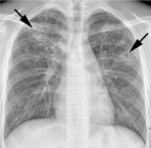
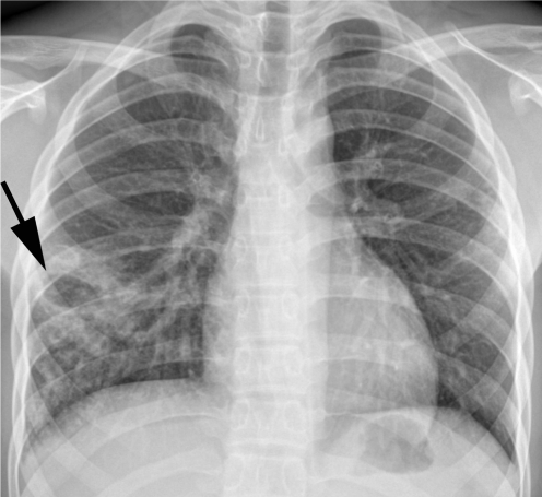
Results: Of 58 patients, three (5%) had normal chest radiographs. Cavitary lesions were present in 25 (45%) of 55 students. Lesions with upper lung zone predominance were observed in 27 (49%) patients, whereas lower lung zone predominance was noted in 18 (33%) patients. The remaining 10 (18%) patients had lesions in both upper and lower lung zones. Pleural effusion was not observed in any patient, nor was the mediastinal lymph node enlargement. Hilar lymph node enlargement was seen in only one (2%) patient. Overall, 37 (67%) students had the typical form of TB, whereas 18 (33%) had TB lesions of the atypical form.
Conclusion: The most common radiographic findings in primary pulmonary TB by recent infection in previously healthy adolescents are upper lung lesions, which were thought to be radiographic findings of reactivation pulmonary TB by remote infection.
Keywords: Adolescent; Mycobacterium tuberculosis; Pulmonary tuberculosis; Thoracic radiography.
Figures

References
-
- Diagnostic Standards and Classification of Tuberculosis in Adults and Children. This official statement of the American Thoracic Society and the Centers for Disease Control and Prevention was adopted by the ATS Board of Directors, July 1999. This statement was endorsed by the Council of the Infectious Disease Society of America, September 1999. Am J Respir Crit Care Med. 2000;161:1376–1395. - PubMed
-
- Small PM, Fujiwara PI. Management of tuberculosis in the United States. N Engl J Med. 2001;345:189–200. - PubMed
-
- Lee KS, Song KS, Lim TH, Kim PN, Kim IY, Lee BH. Adult-onset pulmonary tuberculosis: findings on chest radiographs and CT scans. AJR Am J Roentgenol. 1993;160:753–758. - PubMed
-
- Lee JY, Lee KS, Jung KJ, Han J, Kwon OJ, Kim J, et al. Pulmonary tuberculosis: CT and pathologic correlation. J Comput Assist Tomogr. 2000;24:691–698. - PubMed
-
- Jeong YJ, Lee KS. Pulmonary tuberculosis: up-to-date imaging and management. AJR Am J Roentgenol. 2008;191:834–844. - PubMed
MeSH terms
LinkOut - more resources
Full Text Sources
