

Value of power Doppler and gray-scale US in the diagnosis of carpal tunnel syndrome: contribution of cross-sectional area just before the tunnel inlet as compared with the cross-sectional area at the tunnel
- PMID: 21076589
- PMCID: PMC2974225
- DOI: 10.3348/kjr.2010.11.6.632
Value of power Doppler and gray-scale US in the diagnosis of carpal tunnel syndrome: contribution of cross-sectional area just before the tunnel inlet as compared with the cross-sectional area at the tunnel
Abstract
Objective: To determine the value of gray-scale and power Doppler ultrasonography in the evaluation of carpal tunnel syndrome (CTS).
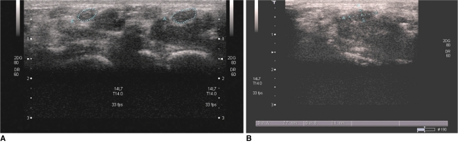
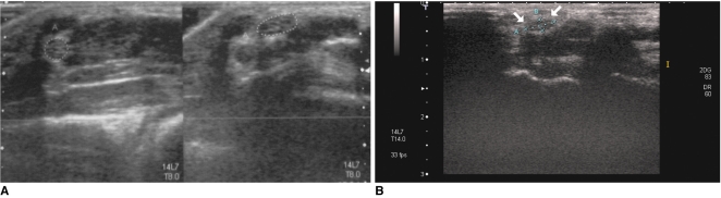
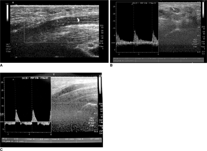
Materials and methods: Median nerves at the carpal tunnel were evaluated by using gray-scale and power Doppler ultrasonography and by using accepted and new criteria in 42 patients with CTS (62 wrists) confirmed by electromyogram and 33 control subjects. We evaluated the cross-sectional area of the nerve just proximal to the tunnel inlet (CSAa), and at mid level (CSAb). We then calculated the percentage area increase of CSAb, and area difference (CSAb-CSAa). We measured two dimensions of the nerve at the distal level to calculate the flattening ratio. The power Doppler ultrasonography was used to assess the number of vessels, which proceeded to give a score according to the vessel number, and lastly evaluated the statistical significance by comparing the means of patients with control subjects by the Student t test for independent samples. Sensitivities and specificities were determined for sonographic characteristics mentioned above. We obtained the receiver operating characteristic (ROC) curve to assess the optimal cut-off values for the diagnosis of CTS.
Results: A statistically significant difference was found between patients and the control group for mean CSAb, area difference, percentage area increase, and flattening ratio (p < 0.001, p < 0.001, p < 0.001, p < 0.05, respectively). From the ROC curve we obtained optimal cut-off values of 11 mm(2) for CSAb, 3.65 for area difference, 50% for the percentage of area increase, and 2.6 for the flattening ratio. The mean number of vessels obtained by power Doppler ultrasonography from the median nerve was 1.2. We could not detect vessels from healthy volunteers. Mean CSAbs related to vascularity intensity scores were as follows: score 0: 12.3 ± 2.8 mm(2), score 1: 12.3 ± 3.1 mm(2), score 2: 14.95 ± 3.5 mm(2), score 3: 19.3 ± 3.8 mm(2). The mean PI value in vessels of the median nerve was 4.1 ± 1.
Conclusion: Gray-scale and power Doppler ultrasonography are useful in the evaluation of CTS.
Keywords: Carpal tunnel syndrome; Power Doppler; Ultrasonography.
Figures
Comment in
-
Re: value of power Doppler and gray-scale US in the diagnosis of carpal tunnel syndrome: contribution of cross-sectional area just before the tunnel inlet as compared with the cross-sectional area at the tunnel.Korean J Radiol. 2011 Mar-Apr;12(2):267. doi: 10.3348/kjr.2011.12.2.267. Epub 2011 Mar 3. Korean J Radiol. 2011. PMID: 21430948 Free PMC article. No abstract available.
References
-
- Duncan I, Sullivan P, Lomas F. Sonography in the diagnosis of carpal tunnel syndrome. AJR Am J Roentgenol. 1999;173:681–684. - PubMed
-
- Sarría L, Cabada T, Cozcolluela R, Martínez-Berganza T, García S. Carpal tunnel syndrome: usefulness of sonography. Eur Radiol. 2000;10:1920–1925. - PubMed
-
- Chen P, Maklad N, Redwine M, Zelitt D. Dynamic high-resolution sonography of the carpal tunnel. AJR Am J Roentgenol. 1997;168:533–537. - PubMed
-
- Klauser AS, Halpern EJ, De Zordo T, Feuchtner GM, Arora R, Gruber J, et al. Carpal tunnel syndrome assessment with US: value of additional cross-sectional area measurements of the median nerve in patients versus healthy volunteers. Radiology. 2009;250:171–177. - PubMed
-
- Buchberger W, Judmaier W, Birbamer G, Lener M, Schmidauer C. Carpal tunnel syndrome: diagnosis with high-resolution sonography. AJR Am J Roentgenol. 1992;159:793–798. - PubMed
Publication types
MeSH terms
LinkOut - more resources
Full Text Sources
Medical
Research Materials
Miscellaneous
