

Review
doi: 10.3348/kjr.2010.11.6.687.
Epub 2010 Oct 29.
Invasive ductal carcinoma in a mammary hamartoma: case report and review of the literature
Affiliations
- PMID: 21076596
- PMCID: PMC2974232
- DOI: 10.3348/kjr.2010.11.6.687
Item in Clipboard
Review
Invasive ductal carcinoma in a mammary hamartoma: case report and review of the literature
Korean J Radiol.
2010 Nov-Dec.
Abstract
Mammary hamartomas are typically a benign condition and rarely develop into malignant lesions. Only 14 cases of carcinomas associated with a hamartoma have been documented in the literature. In this case report, we describe a case of invasive ductal carcinoma within a hamartoma in a 72-year-old woman. Mammography, ultrasonography, and magnetic resonance imaging showed the features of a typical hamartoma with a suspicious mass arising in it. This case illustrates the importance of identification of unusual findings in a typical mammary hamartoma on radiologic examinations.
Keywords: Invasive ductal carcinoma; Mammary hamartoma.
Figures
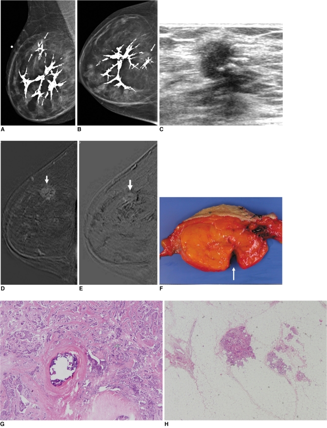
Invasive ductal carcinoma and mammary hamartoma in 72-year-old woman. A, B. Mediolateral oblique and craniocaudal views of right breast show fat-containing mass including dystrophic calcifications, suggesting hamartoma. There is focal asymmetry (arrows) at 12 o'clock within hamartoma. C. US of right breast at 12 o'clock shows spiculated, nonparallel, hypoechoic mass, which corresponds with focal asymmetry on mammogram (A, B). D, E. Standard subtraction image (D) and reverse subtraction image (E) of dynamic MRI show suspicious mass (arrows) within hamartoma, which was early enhanced (D) and washout (E). F. Gross specimen shows 9-cm smooth circumscribed fatty mass. Suspicious mass was excised from fatty mass (arrow). G, H. Photomicrograph of histopathologic specimen of excised suspicious mass reveals invasive ductal carcinoma and dystrophic calcifications (in H) located within carcinoma.
References
-
- Stavros AT. Breast ultrasound. Philadelphia: LWW; 2004. pp. 560–569.
-
- Mester J, Simmons RM, Vazquez MF, Rosenblatt R. In situ and infiltrating ductal carcinoma arising in a breast hamartoma. AJR Am J Roentgenol. 2000;175:64–66. - PubMed
-
- Mendiola H, Henrik-Nielsen R, Dyreborg U, Blichert-Toft M, Al-Hariri JA. Lobular carcinoma in situ occurring in adenolipoma of the breast. Report of a case. Acta Radiol Diagn (Stockh) 1982;23:503–505. - PubMed
-
- Baron M, Ladonne JM, Gravier A, Picquenot JM, Berry M. Invasive lobular carcinoma in a breast hamartoma. Breast J. 2003;9:246–248. - PubMed
Publication types
MeSH terms
LinkOut - more resources
Full Text Sources
Medical
