

Formation of ZnO Micro-Flowers Prepared via Solution Process and their Antibacterial Activity
- PMID: 21076675
- PMCID: PMC2956051
- DOI: 10.1007/s11671-010-9694-y
Formation of ZnO Micro-Flowers Prepared via Solution Process and their Antibacterial Activity
Abstract
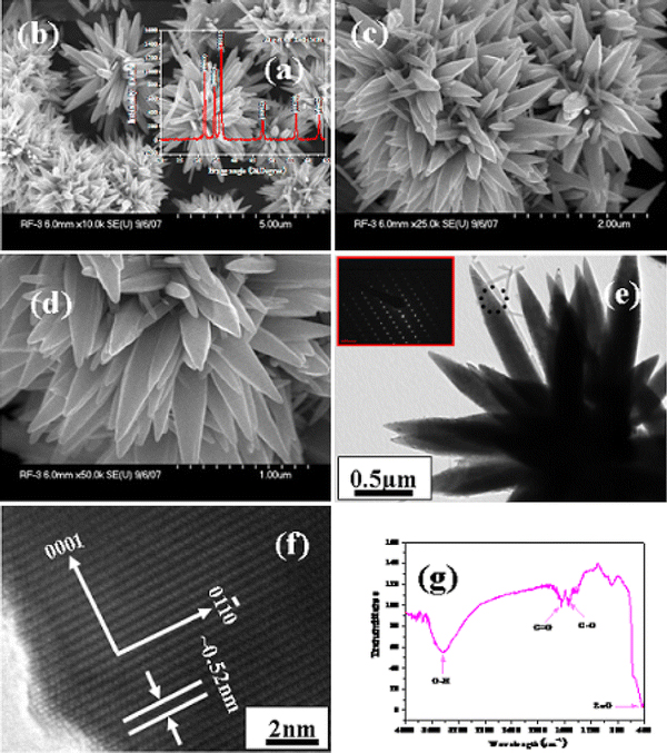
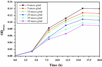
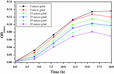
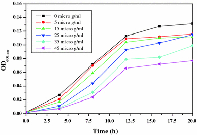
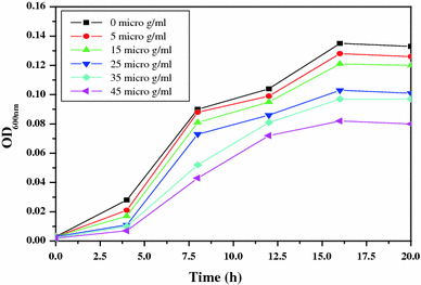
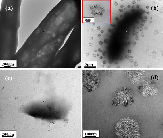
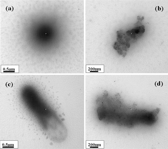
This paper presents the fabrication and characterization of zinc oxide micro-flowers and their antibacterial activity. The micro-flowers of zinc oxide composed of hexagonal nanorods have been prepared via solution process using precursor zinc acetate di-hydrate and sodium hydroxide in 3 h of refluxing time at ~90°C. The antibacterial activities of grown micro-flowers were investigated against four pathogenic bacteria namely S. aureus, E. coli, S. typhimurium and K. pneumoniae by taking five different concentrations (5-45 μg/ml) of ZnO micro-flowers (ZnO-MFs). Our investigation reveals that at lowest concentration of ZnO-MFs solution inhibiting the growth of microbial strain which was found to be 5 μg/ml for all the tested pathogens. Additionally, on the basis of morphological and chemical observations, a chemical reaction mechanism of ZnO-MFs composed of hexagonal nanorods was also proposed.
Figures
References
-
- L.E. Hancook, M.S.Gilmore, V.A. Fischetti, R.P.Novick, J.J. Ferretti, D.A. Portnoy, J.I. Rood (eds.), editor. ASM Press, Washington DC; 2000.
-
- Holister P, Weener JW, Romas CV, Harper T. Nanoparticles. Technology white papers 3. Scientific Ltd, London; 2003.
-
- Ciofi N, Torsi L, Ditaranto N, Sabatini L, Zambonin PG, Tantillo G, Ghibelli L, Alessio MD, Bleve-Zacheo T, Traversa E. Appl. Phys. Lett. 2004. p. 2417. Bibcode number [2004ApPhL..85.2417C] - DOI
LinkOut - more resources
Full Text Sources
