

Case Reports
doi: 10.1159/000320246.
Conjunctival Melanoma: A New Clinical and Therapeutical Approach
Affiliations
- PMID: 21076688
- PMCID: PMC2978741
- DOI: 10.1159/000320246
Item in Clipboard
Case Reports
Conjunctival Melanoma: A New Clinical and Therapeutical Approach
Case Rep Dermatol.
.
Abstract
Melanoma involving the conjunctiva is extremely rare. Graver prognosis has been reported with primary conjunctival melanoma than with their cutaneous counterparts [Collin et al.: Aust N Z J Ophthalmol 1986;14:29-34]. Among conjunctival melanomas, two significant risk factors for tumour-related death have been identified: (i) age older than 55 years and (ii) unfavourable tumour location (caruncle, cornea, fornix, palpebral conjunctiva) [Werschnik and Lommatzsch: Am J Clin Oncol 2002;25:248-255]. Here we present a rare case of lentigo maligna involving the palpebral, bulbar conjunctiva and the caruncle. We describe dermoscopic patterns observed and the use of a novel ocular melanoma therapy with topical imiquimod.
Figures
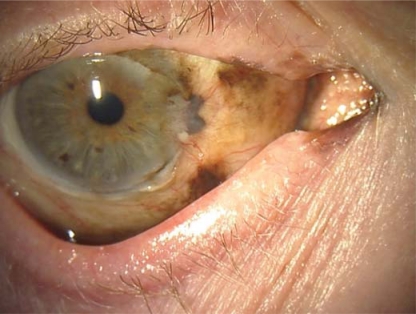
Several ill-defined, millimetric brown and black macules located in the bulbar, palpebral and tarsal conjunctiva and the caruncle could be observed.

Light and dark brown globules and dots could be observed in a parallel pattern. Brown blotches and arborizing vessels in the palpebral and bulbar conjunctiva were also found.

Red and gray colours were present mostly in the tarsal conjunctiva. Vessels with a rope-ladder pattern (scar-like) were found.

Histopathological examination revealed atypical proliferation of melanocytes in a lentiginous pattern. Melanocytes showed a pagetoid distribution between the keratinocytes in the epithelium.

Melanocytic marker Melan-A expression was consistent with LM in immunohistochemical studies.

One-year follow-up showed the patient was disease-free.
References
-
- Collin JR, Garner A, Allen LH, Hungerford JL. Malignant melanoma of the eyelid and conjunctiva. Aust N Z J Ophtalmol. 1986;14:29–34. - PubMed
-
- Werschnik C, Lommatzsch PK. Long-term follow-up of patients with conjunctival melanoma. Am J Clin Oncol. 2002;25:248–255. - PubMed
-
- Saornil MA, Becerra E, Méndez MC, Blanco G. Conjunctival tumors. Arch Soc Esp Oftalmol. 2009;84:7–22. - PubMed
-
- Shields Cl, Shields JA. Ocular melanoma: relatively rare but requiring respect. Clin Dermatol. 2009;27:122–133. - PubMed
-
- Grin JM, Grant-Kels JM, Grin CM, Berke A, Kels BD. Ocular melanomas and melanocytic lesions of the eye. J Am Acad Dermatol. 1998;38:716–730. - PubMed
Publication types
LinkOut - more resources
Full Text Sources
