Review
. 2011 Mar;68(6):991-1003.
doi: 10.1007/s00018-010-0588-z. Epub 2010 Nov 13.

Translation initiation: variations in the mechanism can be anticipated

Affiliations
Review

Translation initiation: variations in the mechanism can be anticipated

Naglis Malys et al. Cell Mol Life Sci. 2011 Mar.

Abstract

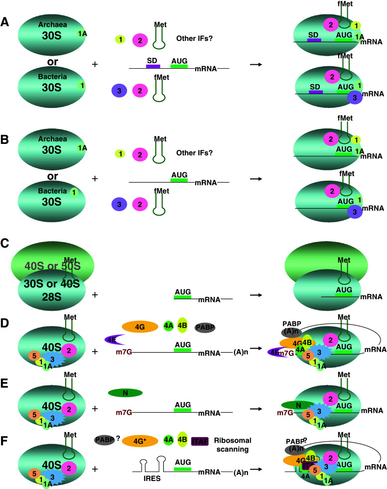
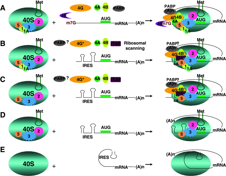
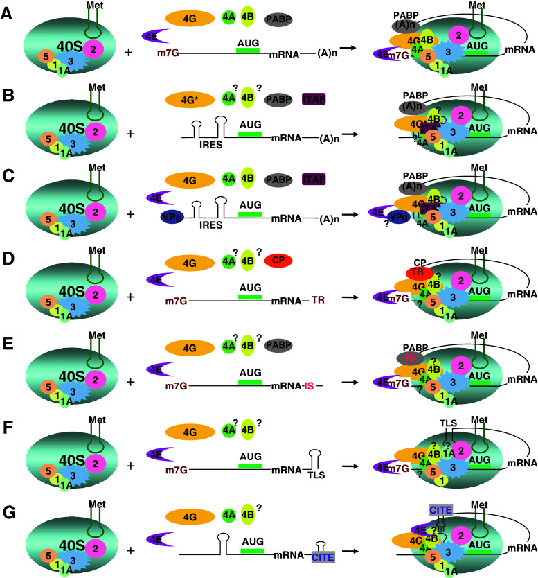
Translation initiation is a critical step in protein synthesis. Previously, two major mechanisms of initiation were considered as essential: prokaryotic, based on SD interaction; and eukaryotic, requiring cap structure and ribosomal scanning. Although discovered decades ago, cap-independent translation has recently been acknowledged as a widely spread mechanism in viruses, which may take place in some cellular mRNA translations. Moreover, it has become evident that translation can be initiated on the leaderless mRNA in all three domains of life. New findings demonstrate that other distinguishable types of initiation exist, including SD-independent in Bacteria and Archaea, and various modifications of 5' end-dependent and internal initiation mechanisms in Eukarya. Since translation initiation has developed through the loss, acquisition, and modification of functional elements, all of which have been elevated by competition with viral translation in a large number of organisms of different complexity, more variation in initiation mechanisms can be anticipated.


Figures

Fig. 1
Fig. 1
Comparison of key translation initiation mechanisms. Figure shows simplified diagrams of the ribosome recruitment process up to the stage of AUG recognition. Ribosomal subunit initiation factors and mRNA with key structural elements are depicted. The following translation initiation mechanisms are shown: a SD-dependent, b SD-independent; both (a, b) are common to Archaea and Bacteria; c a leaderless mechanism, which occurs in Archaea, Bacteria, Eukarya, and mitochondria and may be initiated by small ribosomal subunits or 70S and 80S ribosomes; d 5′ end-dependent, which is represented as a ‘canonical’ cap-dependent mechanism; e 5′ end-dependent mechanism found in Hantavirus, which uses a viral multifunctional translation factor that substitutes cap binding complex; f internal initiation, which is represented as an IRES-driven mechanism. Initiation factors aIF1, aIF1A, and eIF2 of Archaea are labeled as 1, 1A, and 2; IF1, IF2, and IF3 of Bacteria are 1, 2, and 3; eIF1, eIF1A, eIF2, eIF3, eIF4A, eIF4B, eIF4E, eIF4G, and eIF5 of Eukarya are 1, 1A, 2, 3, 4A, 4B, 4E, 4G, and 5, respectively
Fig. 2
Fig. 2
Variations of IRES types in the viral translation initiation. The diagram is a schematic representation of the ribosomal subunits, IFs, and mRNA involved in the IRES-driven translation. Highlighted mechanisms are adopted from [33, 82]. Canonical initiation requires the 5′capped mRNA and all IFs (a), whereas IRES-driven initiation uses a broad variety of IRESs and different subsets of IFs (be). b, c are found in Picornaviridae and require ITAFs. b IRES-driven initiation involving the majority of canonical eIFs except eIF4E and ribosomal scanning that occurs prior to the start codon is reached (e.g., poliovirus (PV), classed as type 1 IRES by [33]). c The initiation codon is located immediately after the 3′ end of IRES. Here, ribosomal scanning does not occur, and eIF1 and eIF1A are not required [e.g., encephalomyocarditis virus (EMCV) [86, 87] (type 2)]. d Hepatitis C virus (HCV, Hepacivirus genus, Flaviviridae) IRES directly attaches Hepatitis C virus ribosomal subunit to the initiation codon, and limited eIFs are required (type 3). e Initiation in CrPV (Cripavirus genus, Dicistroviridae) that has adapted IRES, which mimics a met-tRNA, and translation initiation (type 4) does occur. All initiation factors are labeled as in Fig. 1. 4G*, truncated eIF4G
Fig. 3
Fig. 3
Variations in the translation initiation mechanisms for plus-strand RNA viruses. The diagram depicts relatively well-established cases in virus families or genera, and does not represent in detail variations that occur inside the virus family or genus (not all factors are shown; question marks indicate unknown or uncertain details). a Initial 43S subunit recruitment step for the canonical cap-dependent translation. Diagrams (bg) illustrate variations of initiation factors and structural elements that are involved in the initial initiation step for viral mRNAs that lack a 7-methyguanosine cap, poly(A) tail, or both: b translation is based on internal initiation via IRES (e.g., PV or EMCV); c initiation involves IRES and viral protein VPg that is covalently linked to the genomic RNA and interacts with eIF4E and eIFiso4E, but its role in translation initiation has not been fully understood [e.g., tobacco etch virus (TEV, Potyviridae) and feline calicivirus (FCV, Caliciviridae)]; d viral coat protein (CP) evicts the PABP and stimulates translation by interacting with eIF4G and 3′ terminal RNA region (TR) of the nonpolyadenylated viral RNA [e.g., alfalfa mosaic virus (AMV) from Bromoviridae]; e PABP stimulates translation by interacting with the internal sequence (IS) in the 3′ end of the nonpolyadenylated RNA [e.g., dengue virus (DENV) from Flavivirus genus Flaviviridae]; f initiation involves the tRNA-like structure (TLS) in the 3′ end of viral RNA [e.g., turnip yellow mosaic virus (TYMV) from Tymovirus genus (Tymoviridae)]; g noncapped and nonpolyadenylated RNA translation is initiated via 3′CITE [e.g., barley yellow dwarf virus (BYDV) from Luteovirus genus (Luteoviridae) and tobacco necrosis virus (TNV) from Necrovirus genus (Tombusviridae)]. Factors eIF4E or eIF-iso4E, and eIF4G or eIF-iso4G are shown under 4E and 4G, respectively. All other initiation factors are labeled as in Fig. 1

Similar articles

Cited by

References

    1. Londei P. Translation. In: Cavicchioli R, editor. Archaea: cellular and molecular biology. Washington: ASM Press; 2007. pp. 175–208.
    1. Kozak M. Initiation of translation in prokaryotes and eukaryotes. Gene. 1999;234:187–208. doi: 10.1016/S0378-1119(99)00210-3. - DOI - PubMed
    1. Woese C, Kandler O, Wheelis M. Towards a natural system of organisms: proposal for the domains Archaea, Bacteria, and Eucarya. Proc Natl Acad Sci USA. 1990;87:4576–4579. doi: 10.1073/pnas.87.12.4576. - DOI - PMC - PubMed
    1. Kyrpides NC, Woese CR. Universally conserved translation initiation factors. Proc Natl Acad Sci USA. 1998;95:224–228. doi: 10.1073/pnas.95.1.224. - DOI - PMC - PubMed
    1. Spahn CMT, Beckmann R, Eswar N, Penczek PA, Sali A, Blobel G, Frank J. Structure of the 80S ribosome from Saccharomyces cerevisiae -tRNA-ribosome and subunit–subunit interactions. Cell. 2001;107:373–386. doi: 10.1016/S0092-8674(01)00539-6. - DOI - PubMed

Publication types

MeSH terms

LinkOut - more resources