. 2011 Feb 18;6(2):192-7.
doi: 10.1021/cb100323z. Epub 2010 Nov 24.

Cardiac induction of embryonic stem cells by a small molecule inhibitor of Wnt/β-catenin signaling

Affiliations

Cardiac induction of embryonic stem cells by a small molecule inhibitor of Wnt/β-catenin signaling

Hanmin Wang et al. ACS Chem Biol. .

Abstract

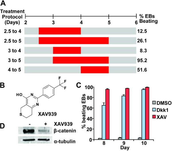
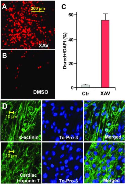
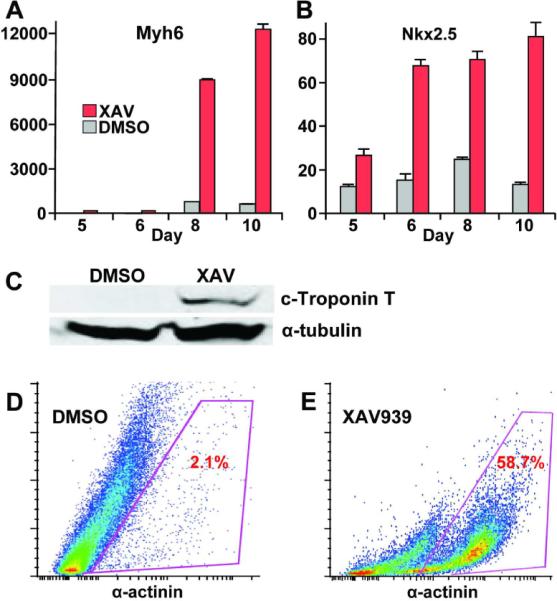
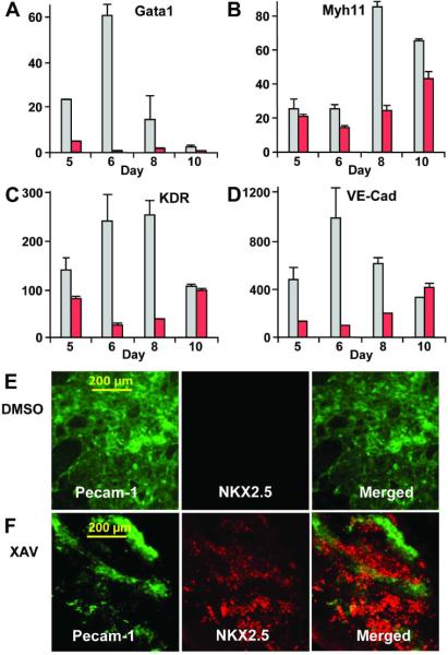
In vitro differentiation of embryonic stem cells is tightly regulated by the same key signaling pathways that control pattern formation during embryogenesis. Small molecules that selectively target these developmental pathways, including Wnt, and BMP signaling may be valuable for directing differentiation of pluripotent stem cells toward many desired tissue types, but to date only few such compounds have been shown to promote cardiac differentiation. Here, we show that XAV939, a recently discovered small molecule inhibitor of Wnt/β-catenin signaling, can robustly induce cardiomyogenesis in mouse ES cells. Our results suggest that a timely administration of XAV939 immediately following the formation of mesoderm progenitor cells promotes cardiomyogenic development at the expense of other mesoderm derived lineages, including the endothelial, smooth muscle, and hematopoietic lineages. Given the critical role that Wnt/β-catenin signaling plays in many aspects of embryogenesis and tissue regeneration, XAV939 is a valuable chemical probe to dissect in vitro differentiation of stem cells and to explore their regenerative potential in a variety of contexts.


Figures

Figure 1
Figure 1. Inhibition of Wnt/β-catenin signaling with the small molecule XAV939 promotes formation of spontaneously beating embryoid bodies
(A) Critical time window for ES cell cardiomyocytes induction with XAV939 (XAV). XAV treatments from Day 2 to 4, Day 2.5 to 5, Day 3 to 4, Day 3 to 5, and Day 4 to 5 were represented by red horizontal bars and the percentages of embryoid bodies (EBs) that beat spontaneously at day 10 of differentiation are shown on the right. Results were obtained from at least 48 EBs for each time point. (B) Chemical structure of XAV939 (3,5,7,8-Tetrahydro-2-[4-(trifluoromethyl)phenyl]-4H-thiopyrano[4,3-d]pyrimidin-4-one). (C) When administered during the day 3 to 5 window, both DKK1 and XAV939 greatly promoted formation of spontaneously beating EBs in comparison to DMSO (P=0.001 for both, P-value was calculated using a two-tailed Student's t test with paired samples throughout this paper, unless otherwise indicated). Results were obtained from at least 48 EBs in 3 independent experiments and standard error was used for graphic plotting throughout this paper, unless otherwise indicated. (D) Western Blot confirmed that β-catenin level was effectively down-regulated in XAV treated EBs (day 5 samples).
Figure 2
Figure 2. ES cells treated with XAV939 (XAV) from day 3 to 5 formed large areas consisting of spontaneously beating cardiomyocytes
CGR-DsRed ES cells, which express DsRed-Nuc fluorescent protein under the control of cardiac α-MHC promoter, formed numerous red fluorescent nuclei following XAV treatment (A), versus relative few fluorescent nuclei following DMSO treatment (B). (C) Approximately 55.6% of DAPI+ nuclei co-expressed DsRed following XAV treatment, versus 1.8% following DMSO treatment (P=0.009). Results were obtained from 6 fields of XAV-treated EBs (on average, 59.6 DsRed+ cells out of 107.2 DAPI+ cells), and 6 fields of DMSO-treated controls (on average, 1.6 DsRed+ cells out of 92.2 DAPI+). (D) XAV939 treated ES cells formed larger areas of cardiomyocytes that expressed sarcomere proteins α-actinin (above) and cardiac Troponin-T (below). Left panels, immunostaining for sarcomere proteins (green). Middle panels, DAPI stained nuclei (blue). Right panels, merged images. Confocal images were taken using a Zeiss inverted LSM 510 confocal microscope (40×).
Figure 3
Figure 3. XAV939 treatment from day 3 to 5 of ES cell differentiation strongly induces cardiomyogenesis
(A, B) XAV treatment led to huge increase in expression of cardiac markers Myh6 and Nkx2.5. Q-PCR results represent relative expression normalized to that of DMSO-treated cells at Day 0. Measurements were obtained from at least three independent experiments for each time-point. Red bars, XAV939-treated. Gray bars, DMSO-vehicle treated. P-values for Myh6 expression at day 8 and 10 following XAV treatment were both <0.0001, in comparison to DMSO-vehicle treatment as negative controls. P-values for Nkx2.5 expression at day 8 and 10 following XAV treatment were 0.0004 and 0006, respectively, in comparison to DMSO treatment. (C) Western blot showing much higher levels of the cardiac Troponin T protein in XAV-treated ES cells on day 10 in comparison to DMSO-treated controls. Antibody against α-tubulin was used as loading control. (E) Representative FACS analysis showing an approximately 28-fold increase in the fraction of α-actinin+ cells following XAV939 treatment vs. DMSO controls.
Figure 4
Figure 4. XAV939 treatment promotes cardiomyogenesis at the expense of other mesoderm lineages
XAV939 treatment decreased the expressions of mesoderm-derived cell lineage markers, including the hematopoietic marker Gata1 (A), the smooth muscle-specific myosin heavy chain gene Myh11 (B), and the endothelial markers Flk-1 and vascular endothelium-cadherin (VE-cad) (C, D). XAV treatment led to huge increase in expression of cardiac markers Myh6 and Nkx2.5. Q-PCR results represent relative expression normalized to that of DMSO-treated cells at Day 0. Measurements were obtained from at least three independent experiments for each time-point. Red bars, XAV939-treated. Gray bars, DMSO-vehicle treated. (E) DMSO-treated ES cells rarely formed cells that express the cardiac Nkx2.5 protein (red immunostaining, right panel), but rather formed large patches of cells that express the endothelial Pecam-1 protein (green immunostaining, middle panel). (F) By contrast, XAV939-treated ES cells formed large areas of Nkx2.5 expressing cardiomyocytes (left panel), and considerably smaller areas of Pecam-1 expressing endothelial cells (middle panel). (E, F) Right panels, merged images.

Similar articles

Cited by

References

    1. Fukuda K, Yuasa S. Stem cells as a source of regenerative cardiomyocytes. Circulation research. 2006;98:1002–1013. - PubMed
    1. Laflamme MA, Chen KY, Naumova AV, Muskheli V, Fugate JA, Dupras SK, Reinecke H, Xu C, Hassanipour M, Police S, O'Sullivan C, Collins L, Chen Y, Minami E, Gill EA, Ueno S, Yuan C, Gold J, Murry CE. Cardiomyocytes derived from human embryonic stem cells in pro-survival factors enhance function of infarcted rat hearts. Nature biotechnology. 2007;25:1015–1024. - PubMed
    1. Okita K, Ichisaka T, Yamanaka S. Generation of germline-competent induced pluripotent stem cells. Nature. 2007;448:313–317. - PubMed
    1. Okita K, Nakagawa M, Hyenjong H, Ichisaka T, Yamanaka S. Generation of mouse induced pluripotent stem cells without viral vectors. Science. 2008;322:949–953. - PubMed
    1. Takahashi K, Tanabe K, Ohnuki M, Narita M, Ichisaka T, Tomoda K, Yamanaka S. Induction of pluripotent stem cells from adult human fibroblasts by defined factors. Cell. 2007;131:861–872. - PubMed

Publication types

MeSH terms