. 2011 Feb;53(2):145-52.
doi: 10.1016/j.cyto.2010.10.007.

Facultative heterochromatin formation at the IL-1 beta promoter in LPS tolerance and sepsis

Affiliations

Facultative heterochromatin formation at the IL-1 beta promoter in LPS tolerance and sepsis

Barbara K Yoza et al. Cytokine. 2011 Feb.

Abstract

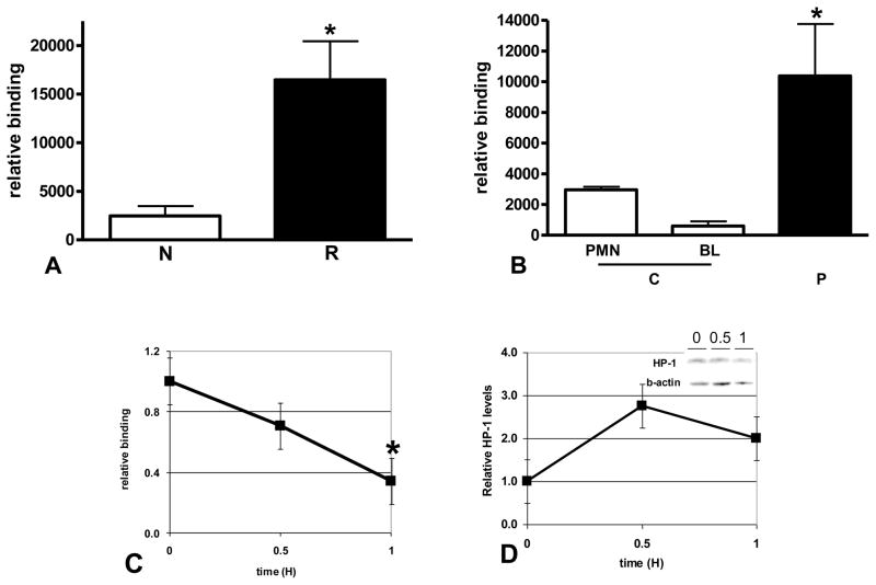
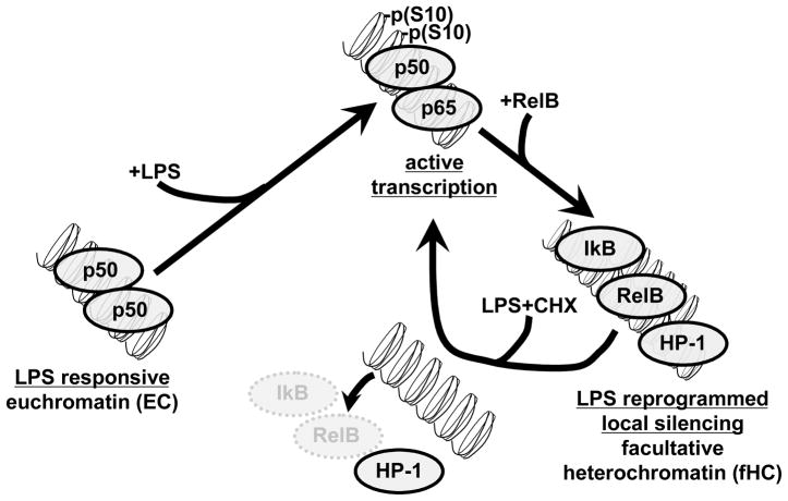
The clinical phenotype in sepsis that is observed as LPS tolerance is determined by silencing of pro-inflammatory genes like IL-1 beta (IL-1β). This study shows that facultative heterochromatin (fHC) silences IL-1β expression during sepsis, where we find dephosphorylated histone H3 serine 10 and increased binding of heterochromatin protein-1 (HP-1) to the promoter. In both human sepsis blood leukocytes and an LPS tolerant human THP-1 cell model, we show that IκBα and v-rel reticuloendotheliosis viral oncogene homolog B (RelB) function as dominant labile mediators of fHC formation at the IL-1β promoter. Protein synthesis inhibition decreases levels of IκBα and RelB, converts silent fHC to euchromatin, and restores IL-1β transcription. We further show TLR dependent NFκB p65 and histone H3 serine 10 phosphorylation binding at the promoter. We conclude that the resolution phase of sepsis, which correlates with survival in humans, may depend on the plasticity of chromatin structure as found in fHC.


Figures

FIGURE 1
FIGURE 1
IL-1β expression is increased by protein synthesis inhibition in reprogrammed cells. A. THP-1 cells were treated as indicated. Total RNA was isolated and IL-1β mRNA measured by real time PCR as described in the methods. Data shown are IL-1β mRNA normalized to GAPDH mRNA in each sample and expressed as relative mRNA levels. Untreated normal THP-1 (N) and reprogrammed THP-1 cells (R) have very low levels of IL-1β mRNA. Normal cells show induced levels of IL-1β mRNA when treated with LPS, whereas reprogrammed cells are significantly repressed (*p<0.001, t-test). Protein synthesis inhibition with CHX reverses IL-1β repression (solid bars) in reprogrammed cells. There is a significant difference (**p<0.05) between LPS alone and CHX or LPS+CHX treated reprogrammed cells; there is no significant difference (p>0.05) between LPS treated normal cells, CHX treated reprogrammed cells or LPS+CHX treated normal or reprogrammed cells (one-way ANOVA, Bonferroni multicomparison post-test). B. BL were isolated and treated as indicated. Total RNA was isolated and IL-1β mRNA measured by real time PCR as described in the methods. Data shown are IL-1β mRNA normalized to GAPDH mRNA in each sample and expressed as fold increase relative to untreated mRNA levels. Control BL (C) show induced levels of IL-1β mRNA when treated with LPS, whereas SSI patient BL (P) are significantly repressed (*p<0.005, t-test) that is reversed by protein synthesis inhibition (solid bars). There is no significant difference (p>0.05) between LPS treated control BL and LPS+CHX treated SSI patient BL; there is a significant difference (**p<0.05) between LPS alone and LPS+CHX treated patient BL (one-way ANOVA, Bonferroni multicomparison post-test). C. Control PMN IL-1β mRNA levels induced by LPS is shown for comparison. There is no significant difference (p>0.05) in IL-1β mRNA levels induced by LPS in control BL and PMN.
FIGURE 2
FIGURE 2
IκB and RelB binding to the IL-1β promoter are increased in reprogrammed cells. A. ChIP assays were used to measure IκB and RelB binding to the IL-1β promoter in normal (N) and reprogrammed (R) THP-1 cells as described in the methods. A significant increase in binding was found for both proteins in reprogrammed cells (*p<0.01, t-test). Data shown are normalized to input DNA and expressed as fold increase relative to binding in normal cells. B. Reprogrammed cells were transfected with IκB siRNA and stimulated with LPS. IκB knockdown significantly (*p<0.001) increased IL-1β expression when compared to cells transfected with control siRNA (ctrl siRNA). C and D. ChIP assays were used to measure IκB (B) and RelB (C) binding to the IL-1β promoter in control (C) and SSI patient BL (P) as described in the methods. A significant increase in binding was found for both proteins in SSI patient BL (*p<0.05, one-way ANOVA, Bonferroni multicomparison posttest). Low levels of binding for IκB and RelB to the IL-1β promoter in control PMN is also shown.
FIGURE 3
FIGURE 3
Inhibition of protein synthesis decreases IκB and RelB nuclear protein and promoter binding in reprogrammed cells. Reprogrammed THP-1 cells were treated with LPS+CHX for the indicated times, nuclear extracts prepared, and IκB (A) or RelB (B) protein levels in nuclear extracts (30ug) were quantitated by W. blot as described in the methods. Inhibition of protein synthesis decreases IkB and RelB nuclear protein levels. Results shown are the average of 3 separate experiments. A representative blot is shown (inset). IκB (C) or RelB (D) binding to the IL-1β promoter was measured by ChIP assays in reprogrammed THP-1 cells treated with LPS+CHX. Protein synthesis inhibition significantly (*p<0.05) decreases both IκB and RelB binding to the IL-1β promoter. Results shown are the average of three independent experiments.
FIGURE 4
FIGURE 4
Inhibition of protein synthesis increases LPS induced p65 binding and phosphorylation of histone H3 at the IL-1β promoter in reprogrammed cells. NFκB p65 binding (A, B) and H3 (serine10) phosphorylation (C, D) at the IL-1β promoter was measured by ChIP assay in reprogrammed THP-1 cells treated with CHX alone (A, C) or LPS+CHX (B, D) for the indicated times. Inhibition of protein synthesis is unable to induce p65 or histone phosphorylation in normal cells (dotted line) when compared to reprogrammed cells (solid line). Protein synthesis inhibition restores both p65 binding and histone phosphorylation at the IL-1β promoter in reprogrammed cells (solid line). Results shown are the average of three (A, B, C) and 4 (D) independent experiments.
FIGURE 5
FIGURE 5
Formation of fHC represses IL-1β expression in LPS reprogrammed cells. A. ChIP assays were used to show a significant increase in HP-1 binding to the IL-1β promoter in reprogrammed (R) THP-1 cells (*p<0.02) when compared to normal (N). B. ChIP assays were used to compare HP-1 binding to the IL-1 promoter in control (C) and and SSI patient BL (P). A significant increase in binding was found in SSI patient BL (*p<0.05, one-way ANOVA, Bonferroni multicomparison post-test). Low levels of HP-1 binding to the IL-1β promoter in control PMN is also shown. C. ChIP assays were used to measure HP-1 binding to the IL-1β promoter in reprogrammed THP-1 cells treated with LPS+CHX for the indicated times. Protein synthesis inhibition significantly (*p<0.05) decreases HP-1 binding to the IL-1β promoter. Results shown are the average of three independent experiments. D. Reprogrammed THP-1 cells were treated with LPS+CHX for the indicated times, nuclear extracts prepared, HP-1 protein levels in nuclear extracts were quantitated by W. blot as described in the methods. HP-1 levels are not sensitive to treatment with CHX. Results shown are the average of 4 separate experiments. A representative blot is shown (inset).
FIGURE 6
FIGURE 6
LPS reprogramming and formation of fHC. At the onset of SSI and in LPS treated THP-1 cells, NFκB is activated and transcription of proinflammatory genes like TNFα and IL-1β is observed. Transcription is associated with NFκB p65 binding and histone H3 S10 phosphorylation at the promoter. Induction of these genes is followed by a period of LPS reprogramming that is characterized by repressed transcription, where SSI patient BL and reprogrammed THP-1 cells are no longer responsive to LPS stimulation. Reprogramming is associated with RelB, IκB and HP-1 binding to the promoter. In this study, we show that inhibition of protein synthesis (by addition of CHX) can restore LPS responsiveness in reprogrammed cells. We found that degradation of RelB and IκB is accompanied by release of HP-1 binding to the promoter. p65 binding and histone H3 S10 phosphorylation at the promoter correlated with the increased transcription of IL-1. As depicted in this model, our results suggest that in normal healthy controls and in naïve cells, proinflammatory genes have the molecular features of euchromatin (EC) that are responsive to LPS activation. In SSI and in LPS reprogrammed cells, silenced proinflammatory genes assume features of facultative heterochromatin (fHC). Degradation of the labile transacting factors, RelB and IκB, is accompanied by release of the heterochromatin protein, HP-1. Restoration of LPS response is characterized by binding of p65 and H3 S10 phosphorylation at the IL-1 promoter and increased IL-1 mRNA.

Similar articles

Cited by

References

    1. Hotchkiss RS, Karl IE. The pathophysiology and treatment of sepsis. N Engl J Med. 2003;348:138–50. - PubMed
    1. Bone RC. Why sepsis trials fail. JAMA. 1996;276:565–6. - PubMed
    1. Foster SL, Medzhitov R. Gene-specific control of the TLR-induced inflammatory response. Clin Immunol. 2009;130:7–15. - PMC - PubMed
    1. West MA, Heagy W. Endotoxin tolerance: A review. Crit Care Med. 2002;30:S64–S73. - PubMed
    1. McCall CE, Yoza BK. Gene silencing in severe systemic inflammation. Am J Respir Crit Care Med. 2007;175:763–7. - PMC - PubMed

Publication types

MeSH terms