. 2011 Jan 28;286(4):3033-46.
doi: 10.1074/jbc.M110.165340. Epub 2010 Nov 15.

PLK1 phosphorylates mitotic centromere-associated kinesin and promotes its depolymerase activity

Affiliations

PLK1 phosphorylates mitotic centromere-associated kinesin and promotes its depolymerase activity

Liangyu Zhang et al. J Biol Chem. .

Abstract

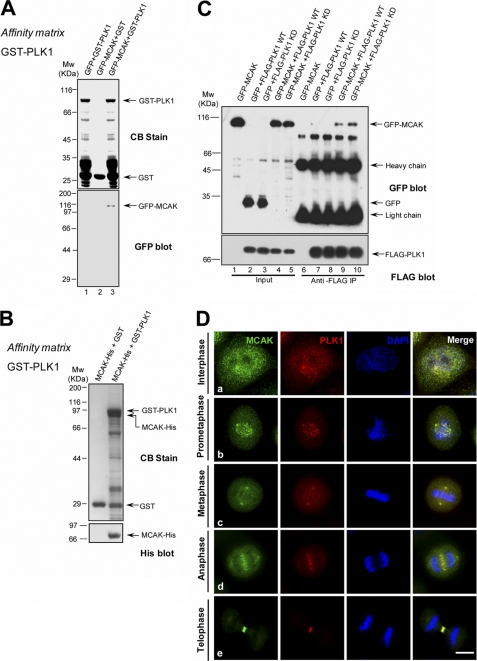
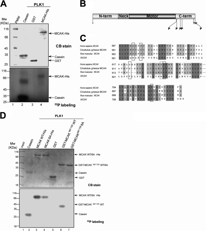
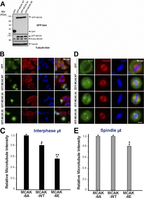
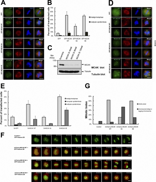
During cell division, interaction between kinetochores and dynamic spindle microtubules governs chromosome movements. The microtubule depolymerase mitotic centromere-associated kinesin (MCAK) is a key regulator of mitotic spindle assembly and dynamics. However, the regulatory mechanisms underlying its depolymerase activity during the cell cycle remain elusive. Here, we showed that PLK1 is a novel regulator of MCAK in mammalian cells. MCAK interacts with PLK1 in vitro and in vivo. The neck and motor domain of MCAK associates with the kinase domain of PLK1. MCAK is a novel substrate of PLK1, and the phosphorylation stimulates its microtubule depolymerization activity of MCAK in vivo. Overexpression of a polo-like kinase 1 phosphomimetic mutant MCAK causes a dramatic increase in misaligned chromosomes and in multipolar spindles in mitotic cells, whereas overexpression of a nonphosphorylatable MCAK mutant results in aberrant anaphase with sister chromatid bridges, suggesting that precise regulation of the MCAK activity by PLK1 phosphorylation is critical for proper microtubule dynamics and essential for the faithful chromosome segregation. We reasoned that dynamic regulation of MCAK phosphorylation by PLK1 is required to orchestrate faithful cell division, whereas the high levels of PLK1 and MCAK activities seen in cancer cells may account for a mechanism underlying the pathogenesis of genomic instability.


Figures

FIGURE 1.
FIGURE 1.
MCAK interacts with PLK1 in vitro and in vivo. A, GST-PLK1 on glutathione beads was incubated with lysates of 293T cells transiently transfected with GFP or GFP-MCAK. The beads were washed and analyzed by Western blotting with an anti-GFP antibody. CB, Coomassie Blue stain. B, GST-PLK1, but not GST, pulled down MCAK-His. GST-PLK1 on glutathione-conjugated beads was incubated with recombinant MCAK-His protein purified from Sf9 cells, followed by Western blot analysis using an anti-His antibody. C, co-immunoprecipitation of FLAG-PLK1 and GFP-MCAK. 293T cells were transiently transfected with GFP-MCAK plus FLAG-PLK1 WT (wild type) or FLAG-PLK1 kinase-dead (K82A). 36 h post-transfection, cells were lysed and incubated with the anti-FLAG antibody coupled to agarose beads. The beads were washed and analyzed by Western blotting with an anti-GFP mouse antibody and then probed with an anti-FLAG antibody. D, co-localization of endogenous MCAK and PLK1 during mitosis. HeLa cells were synchronized to mitosis by a thymidine block release, fixed, and then stained with an anti-MCAK rabbit antibody and an anti-PLK1 mouse antibody. DNA was stained with DAPI. MCAK is labeled in green and PLK1 is in red. Scale bar, 10 μm.
FIGURE 2.
FIGURE 2.
Neck and motor domain of MCAK binds to the kinase domain of PLK1. A and B, GST-PLK1 on glutathione beads was incubated with lysates of 293T cells expressing GFP-MCAK or GFP-MCAK deletion mutants, followed by Western blotting with an anti-GFP antibody. C, GST-PLK1, but not GST, interacts with MBP-MCAK(182–586). GST-PLK1 on glutathione beads was incubated with recombinant MBP-MCAK(182–586) purified from E. coli, followed by Western blot analysis with an anti-maltose-binding protein (MBP) antibody. CB, Coomassie Brilliant Blue. D, schematic representation and summary of the binding studies for a series of MCAK deletion mutants assayed in A–C. +, positive; +/−, weak positive; −, negative. Numbers indicate positions of the amino acid residues. E, schematic illustration of PLK1 functional domain and deletion mutants used in F. Numbers indicate the positions of the amino acid residues. F, GST-PLK1 deletion mutants interact with MCAK-His. GST fusion proteins containing N- and C-terminal PLK bound to glutathione beads were incubated with recombinant MCAK-His purified from Sf9 cells followed by Western blot analysis with an anti-His antibody.
FIGURE 3.
FIGURE 3.
PLK1 kinase phosphorylates MCAK protein in vitro. A, bacterially recombinant MCAK-His and GST were incubated with insect cell recombinant PLK1 kinase in an in vitro phosphorylation reaction as described under “Materials and Methods.” Samples were separated by SDS-PAGE and stained with Coomassie Brilliant Blue (CB) to ensure equal amounts of MCAK-His and GST used in the reactions. The gels were then dried and exposed to an x-ray film. B, schematic illustration of sites of MCAK phosphorylated by PLK1. Recombinant MCAK-His and GST-MCAK(587–725) were phosphorylated by PLK1 in vitro in the absence of [γ-32P]ATP as described under “Materials and Methods,” and phosphorylation sites on MCAK were identified by mass spectrometry. Five phosphorylation sites (Ser592, Ser595, Ser621, Ser633, and Ser715) identified were shown with the boldface P. C, alignment of sequences around the phosphorylation sites for MCAK from human, Chinese hamster (CHO), mouse, and African Clawed Toad (Xenopus). Dark and light shading indicate the identical and conserved residues, respectively. The residues in the black squares indicate the phosphorylation sites. Although the residue Ser632 in the dotted square was not identified by mass spectrometry, it was also mutated in the following experiments as it is in the vicinity of the phosphorylation site Ser633. The numbers indicate the amino acid positions. D, recombinant MCAK-His (WT and its mutants) and GST-MCAK deletion mutants were phosphorylated by PLK1 in vitro as described in A.
FIGURE 4.
FIGURE 4.
PLK1 phosphorylation stimulates the MT depolymerase activity of MCAK. A, expression levels of GFP-MCAK and its phosphorylation site mutants in HeLa cells. HeLa cells were transfected with GFP, GFP-MCAK WT, GFP-MCAK 6A, or GFP-MCAK 6E. At 36 h post-transfection, cells were harvested, boiled in the SDS-PAGE buffer, and analyzed by Western blotting with an anti-GFP antibody (top panel), anti-MCAK antibody (middle panel), or anti-tubulin antibody (as a loading control, bottom panel). B, in vivo analysis of GFP-MCAK and its phosphorylation site mutants in interphase cells. HeLa cells were transfected with GFP, GFP-MCAK WT, GFP-MCAK 6, or GFP-MCAK 6E. At 36 h post-transfection, cells were fixed with methanol and stained for α-tubulin (red) and DAPI (for DNA, blue), respectively. C, statistical analysis of the relative MT density in B as described under “Materials and Methods.” Data are presented as means ± S.E. and derived from 75 cells from three independent experiments. *, p < 0.05; **, p < 0.01. D, in vivo analysis of GFP-MCAK and its phosphorylation sites mutants in mitotic cells. HeLa cells were transfected with GFP, GFP-MCAK WT, GFP-MCAK 6A, or GFP-MCAK 6E and synchronized to metaphase. At 36 h post-transfection, cells were fixed and stained as described in B. E, statistical analysis of relative spindle MT density in D as described under “Materials and Methods.” Data are presented as means ± S.E. and derived from 33 cells from three independent experiments. *, p < 0.05.
FIGURE 5.
FIGURE 5.
Perturbation of PLK1-mediated MCAK phosphorylation resulted in chromosome segregation errors. A, HeLa cells were transfected with GFP-MCAK WT, GFP-MCAK 6A, or GFP-MCAK 6E and synchronized to metaphase with thymidine and MG132 treatment. At 36 h post-transfection, cells were fixed and stained with an anti-α-tubulin antibody (red) and DAPI (blue) to determine the spindle morphology and chromosome position in metaphase. Scale bar, 10 μm. B, statistical analysis of misaligned chromosomes and multipolar spindles in A. Results are presented as means ± S.E. and from n = 150 cells from three independent experiments. C, suppression of MCAK with an siRNA targeted to 3′-UTR. HeLa cells were transfected with a scramble siRNA or MCAK siRNA and harvested at 48 h post-transfection. Cell extracts were probed with an anti-MCAK antibody (top panel) and anti-tubulin antibody (as a loading control, bottom panel). D, HeLa cells were transfected with 40 nm MCAK siRNA for 48 h and then transfected to express GFP, GFP-MCAK WT, GFP-MCAK 6A, or GFP-MCAK 6E for another 36 h. Cells were synchronized, fixed, and stained with an α-tubulin antibody (red) and DAPI (blue) as in A. Scale bar, 10 μm. E, statistical analysis of misaligned chromosomes and multipolar spindles in C. Results are presented as means ± S.E. and from n = 200 cells from three independent experiments. F, real time imaging of chromosome movement in cells expressing cherry-MCAK WT, cherry-MCAK 6A, and cherry-MCAK 6E. Cells were co-transfected with GFP-histone 2B plus cherry-MCAK WT or its phosphorylation site mutants together. At 36 h post-transfection, cells were observed with a DeltaVision system. G, statistical analysis of mitotic arrest and anaphase disruption in F. At least 20 cells were analyzed from three independent experiments for each construct.
FIGURE 6.
FIGURE 6.
PLK1 phosphorylation regulates MCAK intramolecular interaction. A, MCAK exhibits an intramolecular association in vitro. Recombinant GST-MCAK(1–181) and -(182–586) on glutathione beads were incubated with MCAK-His purified from Sf9 cells as described in Fig. 1B. Anti-MCAK antibody was used in Western blot analysis to determine the intramolecular interaction of MCAK. B, PLK1 phosphorylation promotes the intramolecular association of MCAK in vitro. Recombinant MCAK deletion mutant proteins (GST-MCAK(1–181) and GST-MCAK(182–586)) on glutathione-agarose beads were used as an affinity matrix to isolate MCAK WT-His and its phosphorylation sites mutants from Sf9 cells. Anti-MCAK antibody was used in Western blot analysis to determine the phospho-regulation of the intramolecular interaction of MCAK. C, statistical analysis of the relative binding activity in B. The ordinate indicates the binding ratio of the intensity of MCAK (WT, 6A, or 6E)-His to GST-MCAK (MCAK(1–181) or MCAK(182–586)); the abscissa indicates the corresponding binding assay shown in B.

References

    1. Gadde S., Heald R. (2004) Curr. Biol. 14, R797–R805 - PubMed
    1. Tournebize R., Popov A., Kinoshita K., Ashford A. J., Rybina S., Pozniakovsky A., Mayer T. U., Walczak C. E., Karsenti E., Hyman A. A. (2000) Nat. Cell Biol. 2, 13–19 - PubMed
    1. Desai A., Verma S., Mitchison T. J., Walczak C. E. (1999) Cell 96, 69–78 - PubMed
    1. Moores C. A., Yu M., Guo J., Beraud C., Sakowicz R., Milligan R. A. (2002) Mol. Cell 9, 903–909 - PubMed
    1. Ogawa T., Nitta R., Okada Y., Hirokawa N. (2004) Cell 116, 591–602 - PubMed

Publication types

MeSH terms