

Probing the in vitro mechanism of action of cationic lipid/DNA lipoplexes at a nanometric scale
- PMID: 21078679
- PMCID: PMC3045597
- DOI: 10.1093/nar/gkq921
Probing the in vitro mechanism of action of cationic lipid/DNA lipoplexes at a nanometric scale
Abstract
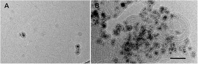
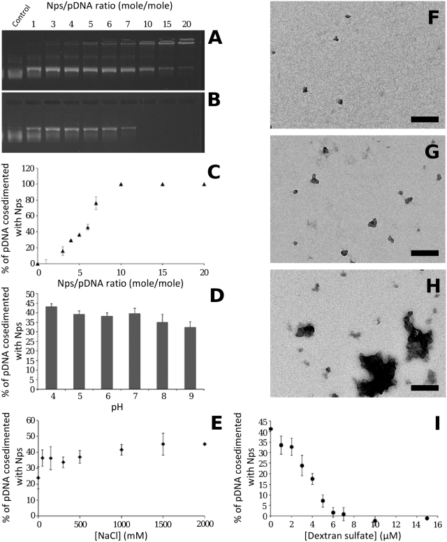
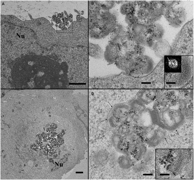
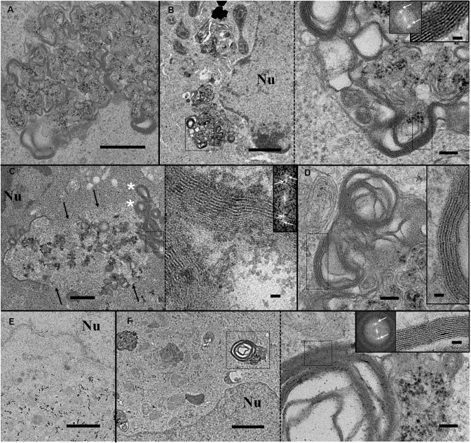
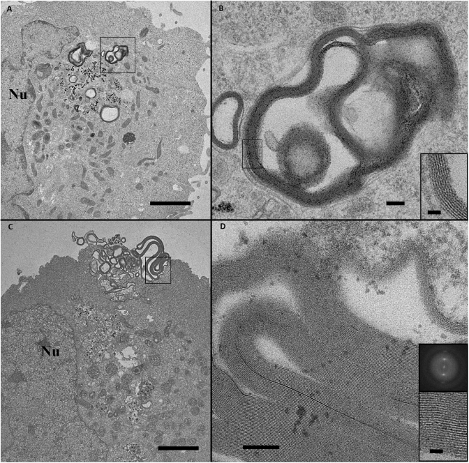
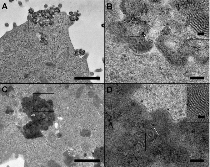
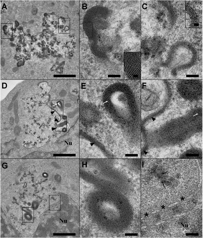
Cationic lipids are used for delivering nucleic acids (lipoplexes) into cells for both therapeutic and biological applications. A better understanding of the identified key-steps, including endocytosis, endosomal escape and nuclear delivery is required for further developments to improve their efficacy. Here, we developed a labelling protocol using aminated nanoparticles as markers for plasmid DNA to examine the intracellular route of lipoplexes in cell lines using transmission electron microscopy. Morphological changes of lipoplexes, membrane reorganizations and endosomal membrane ruptures were observed allowing the understanding of the lipoplex mechanism until the endosomal escape mediated by cationic lipids. The study carried out on two cationic lipids, bis(guanidinium)-tris(2-aminoethyl)amine-cholesterol (BGTC) and dioleyl succinyl paramomycin (DOSP), showed two pathways of endosomal escape that could explain their different transfection efficiencies. For BGTC, a partial or complete dissociation of DNA from cationic lipids occurred before endosomal escape while for DOSP, lipoplexes remained visible within ruptured vesicles suggesting a more direct pathway for DNA release and endosome escape. In addition, the formation of new multilamellar lipid assemblies was noted, which could result from the interaction between cationic lipids and cellular compounds. These results provide new insights into DNA transfer pathways and possible implications of cationic lipids in lipid metabolism.
Figures
Similar articles
-
Brewster angle microscopy and PMIRRAS study of DNA interactions with BGTC, a cationic lipid used for gene transfer.Langmuir. 2008 Sep 2;24(17):9598-606. doi: 10.1021/la703491r. Epub 2008 Jul 30. Langmuir. 2008. PMID: 18665617
-
Nonbilayer phase of lipoplex-membrane mixture determines endosomal escape of genetic cargo and transfection efficiency.Mol Ther. 2005 May;11(5):801-10. doi: 10.1016/j.ymthe.2004.12.018. Mol Ther. 2005. PMID: 15851018
-
Sterically stabilized BGTC-based lipoplexes: structural features and gene transfection into the mouse airways in vivo.J Gene Med. 2001 Sep-Oct;3(5):478-87. doi: 10.1002/jgm.211. J Gene Med. 2001. PMID: 11601761
-
Gene delivery by cationic lipids: in and out of an endosome.Biochem Soc Trans. 2007 Feb;35(Pt 1):68-71. doi: 10.1042/BST0350068. Biochem Soc Trans. 2007. PMID: 17233603 Review.
-
On the mechanism of cationic amphiphile-mediated transfection. To fuse or not to fuse: is that the question?J Membr Biol. 2002 Oct 1;189(3):167-79. doi: 10.1007/s00232-002-1015-7. J Membr Biol. 2002. PMID: 12395282 Review.
Cited by
-
Self-Crosslinking Lipopeptide/DNA/PEGylated Particles: A New Platform for DNA Vaccination Designed for Assembly in Aqueous Solution.Mol Ther Nucleic Acids. 2018 Sep 7;12:504-517. doi: 10.1016/j.omtn.2018.05.025. Epub 2018 Jul 18. Mol Ther Nucleic Acids. 2018. PMID: 30195787 Free PMC article.
-
Critical factors for lentivirus-mediated PRDX4 gene transfer in the HepG2 cell line.Oncol Lett. 2018 Jul;16(1):73-82. doi: 10.3892/ol.2018.8650. Epub 2018 May 7. Oncol Lett. 2018. PMID: 29930713 Free PMC article.
-
Optically-controlled platforms for transfection and single- and sub-cellular surgery.Biophys Rev. 2015 Dec;7(4):379-390. doi: 10.1007/s12551-015-0179-1. Epub 2015 Nov 16. Biophys Rev. 2015. PMID: 28510103 Free PMC article. Review.
-
COVID-19 vaccines: Frequently asked questions and updated answers.Infect Dis Now. 2021 Jun;51(4):319-333. doi: 10.1016/j.idnow.2021.02.007. Epub 2021 Feb 27. Infect Dis Now. 2021. PMID: 33681861 Free PMC article. Review.
-
Cationic liposome/DNA complexes: from structure to interactions with cellular membranes.Eur Biophys J. 2012 Oct;41(10):815-29. doi: 10.1007/s00249-012-0830-8. Epub 2012 Jun 19. Eur Biophys J. 2012. PMID: 22710765 Review.
References
-
- Felgner PL, Barenholz Y, Behr JP, Cheng SH, Cullis P, Huang L, Jessee JA, Seymour L, Szoka F, Thierry AR, et al. Nomenclature for synthetic gene delivery systems. Hum. Gene Ther. 1997;8:511–512. - PubMed
-
- Barteau B, Chèvre R, Letrou-Bonneval E, Labas R, Lambert O, Pitard B. Physicochemical parameters of non-viral vectors that govern transfection efficiency. Curr. Gene Ther. 2008;8:313–323. - PubMed
-
- Khalil IA, Kogure K, Akita H, Harashima H. Uptake pathways and subsequent intracellular trafficking in nonviral gene delivery. Pharmacol. Rev. 2006;58:32–45. - PubMed
-
- Hoekstra D, Rejman J, Wasungu L, Shi F, Zuhorn I. Gene delivery by cationic lipids: in and out of an endosome. Biochem. Soc. Trans. 2007;35:68–71. - PubMed
-
- Midoux P, Breuzard G, Gomez JP, Pichon C. Polymer-based gene delivery: a current review on the uptake and intracellular trafficking of polyplexes. Curr. Gene Ther. 2008;8:335–352. - PubMed
