Review
. 2010 Nov 15;24(22):2480-92.
doi: 10.1101/gad.1973910.

Eph/ephrin molecules--a hub for signaling and endocytosis

Affiliations
Review

Eph/ephrin molecules--a hub for signaling and endocytosis

Mara E Pitulescu et al. Genes Dev. .

Abstract

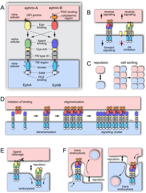
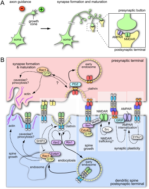
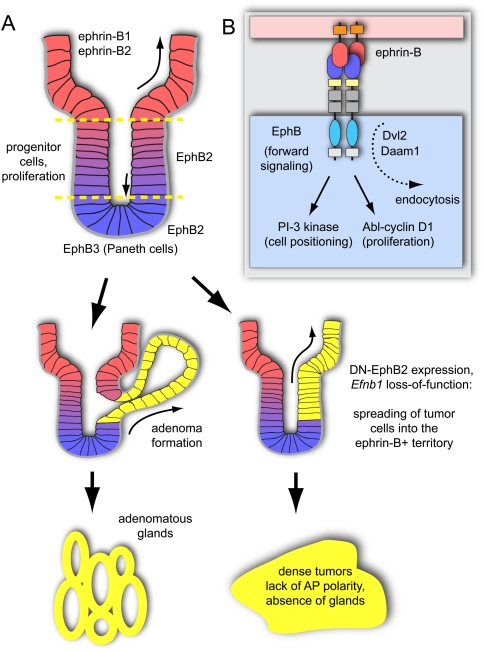
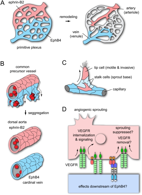
The development, homeostasis, and regeneration of complex organ systems require extensive cell-cell communication to ensure that different cells proliferate, migrate, differentiate, assemble, and function in a coordinated and timely fashion. Eph receptor tyrosine kinases and their ephrin ligands are critical regulators of cell contact-dependent signaling and patterning. Eph/ephrin binding can lead to very diverse biological readouts such as adhesion versus repulsion, or increased versus decreased motility. Accordingly, depending on cell type and context, a limited and conserved set of receptor-ligand interactions is translated into a large variety of downstream signaling processes. Recent evidence indicates that the endocytosis of Eph/ephrin molecules, together with the internalization of various associated tissue-specific effectors, might be one of the key principles responsible for such highly diverse and adaptable biological roles. Here, we summarize recent insights into Eph/ephrin signaling and endocytosis in three biological systems; i.e., the brain, intestine, and vasculature.


Figures

Figure 1.
Figure 1.
Eph/ephrin structure, signaling, and mechanism of action. (A) Domain organization of Eph receptors and ephrin ligands. Cysteine (Cys)-rich, fibronectin (FN) type III, and SAM domains; transmembrane (TM) regions; and tyrosine phosphorylation sites (Y) are indicated. EphA receptors typically bind ephrin-A (GPI-anchored) ligands, and EphB receptors bind ephrin-Bs (arrows). There is limited cross-talk between members of different classes (dashed arrows). (B) Eph/ephrin interactions in trans lead to bidirectional signal transduction. EphA and ephrin-A coexpression in cis impairs receptor activation. (C) Eph/ephrin interactions frequently transduce repulsive signals important for cell migration and cell sorting. (D) Binding Eph/ephrin molecules form heterotetramers to initiate the signal, oligomerize, and further assemble in large receptor clusters that expand laterally trough Eph–Eph cis interactions. (E) Metalloprotease association with the EphA/ephrin-A complex leads to cleavage of the ligand, endocytosis of the complex, and cell–cell repulsion. (F) Eph/ephrin interaction can lead to repulsion also by trans-endocytosis of the complexes in a forward or reverse direction.
Figure 2.
Figure 2.
Eph/ephrin signaling and endocytosis in the nervous system. (A) The growth cones of extending axons are guided by attractive (+) and repulsive (−) cues. Follwing synapse formation and maturation, neurotransmitters are released from the presynaptic side and activate receptors (e.g., AMPAR or NMDAR for excitatory synapses) located in the postsynaptic terminal. (B) In the postsynaptic cell, Eph receptor endocytosis is clathrin-mediated and regulated by the activation of Vav2 (a GEF for Rho family GTPases), Rin1 (a GEF for endosomal Rab5 GTPase), and TIAM1 (a GEF for Rac1 GTPase). Receptor endocytosis and Rac1 GTPase activity are inhibited by the lipid phosphatase SHIP2. Rac1 GTPase (which can get locally activated in endosomes) modifies actin cytoskeleton and has been linked to caveolar internalization or pinocytosis. A similar, Rac1 GTP-dependent mechanism might apply for ephrin-B reverse endocytosis in the presynaptic cell. Linked to forward Eph signaling, Numb (a clathrin adapter) regulates dendritic spine morphogenesis by coupling activated Eph and intersectin (a GEF for Cdc42). Numb regulates not only spine growth, but also synaptic plasticity, probably through Eph-dependent NMDA receptor endocytosis. EphB forward signaling and endocytosis regulates synaptic plasticity by phosphorylation of synaptojanin and enhanced internalization of AMPAR. Postsynaptic ephrin-B reverse signaling leads to GRIP binding and increases AMPAR surface presentation. Some of the mechanisms of endocytosis were derived from studies using soluble Fc fusion proteins and are not validated by cell–cell stimulation experiments.
Figure 3.
Figure 3.
Eph/ephrin signaling regulates intestinal cell positioning and proliferation. (A) EphB2-positive stem and progenitor cells are located near the bottom (lower dashed line) of the intestinal crypts, while ephrin-B1/ephrin-B2-positive differentiated epithelial cells are concentrated at the crypt–villus boundary (upper dashed line). In the small intestine, EphB3-expressing Paneth cells are found at the crypt bottom. Constitutive activation of the Wnt pathway in ApcMin mice leads to epithelial evaginations near the crypt–villus junctions and development of adenomatous polyps (yellow). When Eph/ephrin interactions are compromised, tumor cells spread into the villi, and more advanced colorectal cancers, which lack a glandular structure, develop. (B) Intestinal cell sorting and positioning involves PI3K activity downstream from Eph/ephrin signaling. Cell proliferation is promoted by Abl kinase and cyclin D1. The proteins Dvl and Daam link Eph endocytosis and Wnt signaling.
Figure 4.
Figure 4.
Roles of EphB4 and ephrin-B2 in blood vessel morphogenesis. (A) During vasculogenesis, angioblasts assemble a primitive vascular plexus. Presumptive arterial and venous territories are already marked by complementary expression of ephrin-B2 and EphB4, respectively, before this plexus remodels in arteries (ephrin-B2+), veins (EphB4+), and capillaries. (B) In zebrafish embryos, ventral migration of venous-fated cells from a common precursor vessel leads to segregation of the DA (ephrin-B2+) from the CV (EphB4+). (C) Expression of ephrin-B2 (red) in the leading cells of the sprout (so-called tip cells) partially overlaps with EphB4 (blue) in stalk cells at the sprout base. (D) Ephrin-B2 links the regulation of cell motility and invasiveness to VEGF receptor (VEGFR) endocytosis and signaling. The effects downstream from EphB4 in this context remain unknown.

References

    1. Adams RH, Wilkinson GA, Weiss C, Diella F, Gale NW, Deutsch U, Risau W, Klein R 1999. Roles of ephrinB ligands and EphB receptors in cardiovascular development: Demarcation of arterial/venous domains, vascular morphogenesis, and sprouting angiogenesis. Genes Dev 13: 295–306 - PMC - PubMed
    1. Batlle E, Henderson JT, Beghtel H, van den Born MM, Sancho E, Huls G, Meeldijk J, Robertson J, van de Wetering M, Pawson T, et al. 2002. β-Catenin and TCF mediate cell positioning in the intestinal epithelium by controlling the expression of EphB/ephrinB. Cell 111: 251–263 - PubMed
    1. Batlle E, Bacani J, Begthel H, Jonkheer S, Gregorieff A, van de Born M, Malats N, Sancho E, Boon E, Pawson T, et al. 2005. EphB receptor activity suppresses colorectal cancer progression. Nature 435: 1126–1130 - PubMed
    1. Becker E, Huynh-Do U, Holland S, Pawson T, Daniel TO, Skolnik EY 2000. Nck-interacting Ste20 kinase couples Eph receptors to c-Jun N-terminal kinase and integrin activation. Mol Cell Biol 20: 1537–1545 - PMC - PubMed
    1. Benedito R, Trindade A, Hirashima M, Henrique D, da Costa LL, Rossant J, Gill PS, Duarte A 2008. Loss of Notch signalling induced by Dll4 causes arterial calibre reduction by increasing endothelial cell response to angiogenic stimuli. BMC Dev Biol 8: 117 doi: 10.1186/1471-213X-8-117 - PMC - PubMed

Publication types

MeSH terms

Substances

LinkOut - more resources