. 2011 Jan;31(2):287-99.
doi: 10.1128/MCB.00603-10. Epub 2010 Nov 15.

Expression of type II chorionic gonadotropin genes supports a role in the male reproductive system

Affiliations

Expression of type II chorionic gonadotropin genes supports a role in the male reproductive system

Andrew M Parrott et al. Mol Cell Biol. 2011 Jan.

Abstract

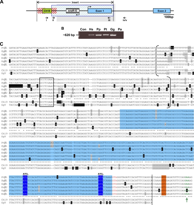
Human chorionic gonadotropin (hCG) is a glycoprotein hormone essential to pregnancy. hCG is heterodimeric and functionally defined by its β subunit. hCGβ evolved from the β subunit of luteinizing hormone in two phases. In the first phase, type I genes (hCGβ3, -5, -7, and -8) acquired changes affecting gene expression and extending the proteins' C terminus. In the second phase, type II genes (hCGβ1 and -2) were formed by the insertion of a DNA element into the type I 5' end. The insertion includes the small noncoding RNA gene snaR-G and has been predicted to drastically change the protein products encoded. We trace the insertion to the common ancestor of the African great apes and show that it contains transcription signals, including snaR-G. Type II transcripts are predominantly expressed in testis. Contrary to predictions, the product of the major mRNA splice form is hCGβ. A novel peptide is encoded by alternatively spliced transcripts. These findings support the view that type II genes evolved in African great apes to function in the male reproductive system.


Figures

FIG. 1.
FIG. 1.
Structure and evolution of type II CGβ genes. (A) Diagram of the LH/CGβ gene cluster on human chromosome 19q13.33. Straight arrows indicate direction of transcription, the large curved arrow denotes inverted segmental duplication of the original CGβ1 gene, and arrowheads depict snaR-G genes. (B) Schematic of LHβ and type I and II CGβ gene structures, illustrating the replacement in type II CGβ genes of a segment common to type I CGβ genes with an insert containing snaR-G (dotted lines). Untranslated regions (UTRs) are depicted as solid boxes; open reading frames (ORFs) are shown as open boxes; introns and promoter regions are shown by solid lines; directions of transcription are shown by bent arrows. The CGβ1/2 first exon is defined here by the first donor splice site (6), and its UTRs (gray boxes) are defined by the first potential ORF. (Adapted from reference with permission of the publisher.)
FIG. 2.
FIG. 2.
Sequence of the type II insert in African great apes. (A) Schematic of the type II CGβ insert (between dotted lines) in the context of CGβ1/2 gene structure (exons in blue). Homology to chromosome 18 (green box), Alu (red hatching), and upstream chromosome 19 sequence (blue hatching) is shown. Bent arrows denote snaR-G and CGβ1/2 transcription directions. PCR forward primers targeting genomic sequence downstream of snaR-G1 or -G2 (i) or snaR-G1 (ii) were coupled with reverse primers targeting hCGβ1/2 exon 1 (iii) and intron 1 (iv) or chimpanzee CGβ1 intron 1 (v). The region shaded gray is aligned in panel C. (B) Ethidium bromide-stained agarose gel of PCR amplification products obtained with primers i and iii, specific for hCGβ1/2 promoter and exon 1, and 293 cell (Hs), bonobo (Pp), chimpanzee (Pt), gorilla (Gg), or orangutan (Pa) genomic DNA or no template (Con). (C) Sequence alignment of African great ape genomic DNA from the CGβ1 and -β2 proximal promoter and first exon (blue shading). snaR-G genes are bracketed, and their B-box promoter is boxed. Upstream chromosome 19 (Ch19) and GgV clone sequences are also aligned, and the limit of their homology is demarcated by the dotted line. Nucleotide heterogeneity is highlighted in gray, and unique bases are highlighted in black. Asterisks denote identity among all sequences except Ch19 and GgV. Putative CGβ1/2 start codons (ATG1 and ATG2) are shaded dark blue, and the start codon of the CGβ antecedent is shaded orange. The new splice donor site (red text) and the consensus splice donor site (green text and arrow) are shown.
FIG. 3.
FIG. 3.
Tissue expression of hCGβ mRNA. (A) Tissue expression of type I mRNA (top panel, 25 cycles; middle panel, 35 cycles) and of type II mRNA (bottom panel, 38 cycles) as determined by RT-PCR. Tissue name abbreviations are defined in Materials and Methods. (B) Comparison of hCGβ1/2 PCR products from positive tissues in panel A. (C) Alternatively spliced hCGβ1/2 mRNAs. The number of clones isolated from each tissue is given. (D) Schematic of the alternative splicing of hCGβ1 and hCGβ2 clones. Nucleotide numbers indicate splice donor (black) or acceptor (gray) sites relative to the transcription start site.
FIG. 4.
FIG. 4.
Type II hCGβ promoter analysis. (A) Schematic of hCGβ1/2 promoter, 5′ UTR, and first exon. The type II insert is demarcated by the dotted line. The extent of hCGβ1/2 sequence included in reporter constructs β1/2L (white bar), β1/2LΔ (hatched bar), β1/2S (black bar), and β1/2SΔ (gray bar) is shown below. (B) Relative luciferase activity in HeLa S3 cells transfected with the β1/2L and β1/2S constructs and their corresponding snaR-G deletion constructs diagrammed in panel A. Activity is normalized to β1L construct, and standard deviations (n = 4 duplicates) are shown. Two-tailed Student's t test: *, P < 0.002, and **, P < 0.001. (C) Northern blots of total RNA from transfected JAr cells. (Top) Cells were transfected with β1L (0, 0.5, 1, or 2 μg) or β1LΔ (2 μg) as indicated, and blots were probed for snaR-G1. (Bottom) As in top panel except that cells were transfected with β2L or β2LΔ and blots were probed for snaR-G2. (D) As in panel C, except that HeLa S3 cells were transfected with 0, 0.3, 0.75, 1.5, or 3 μg of β1L or β2L or with 3 μg of β1LΔ or β2LΔ.
FIG. 5.
FIG. 5.
Type II hCGβ reading frame usage. (A) Schematic of potential hCGβ1a, -2a, and -2b cDNA reading frames. Predicted translation products initiated at ATG1 (black bars) or ATG2 (gray bars) are depicted, and their sizes in amino acids (kDa) are indicated. Exons are depicted as open boxes, and the SanDI restriction site is shown by an arrowhead. (B) Autoradiography of protein products from hCGβ1a, -2a, or -2b cDNA labeled in rabbit reticulocyte (top) or wheat germ (bottom) cell-free transcription/translation systems. Short peptide bands are denoted by the white bracket. (C) Schematic of potential hCGβ1a, -2a, and -2b reading frames. Exons are depicted as open boxes. Reading frames are numbered 1 to 3 with subscript numbers denoting the order of ATG codons relative to the cDNA 5′ end. The first stop codon of each reading frame is numbered in gray. Linkage of the FFL gene to the 5′ part of hCGβ cDNAs is denoted by a dotted line for all F1 to F3 constructs and by a dashed line for the F1′ β2b construct. (D) Immunoblot of extract of HeLa cells transfected with reading frame constructs indicated. Blots were probed with antibodies (Ab) specific for firefly luciferase (FFL; top panel, short exposure; middle panel, longer exposure) and α-tubulin (Tub; bottom panel). (E) Relative luciferase activity in the same extracts as in panel D (n = 4 with duplicate samples).
FIG. 6.
FIG. 6.
Type II hCGβ protein products. (A) Sequence alignment of hCGβ. Key residues are numbered relative to the signal peptide cleavage site (solid line). The Phe residue in the signal peptide sequence (underlined) is replaced by Leu in hCGβ5. Known N- and O-linked glycosylation sites are denoted by gray and black shading, respectively. (B) (Top) Immunoblot of extract of HeLa cells transfected with construct expressing β1a, β2a, or β2b mRNA or empty vector and probed with anti-hCGβ antibody. Extract was incubated in the presence or absence of PNGase F (PNGF) before electrophoresis. (Bottom) Longer exposure of the same immunoblot. An apparent hCGβ1-derived band is denoted by the white arrow. (C) Total RNA extracted from cells in panel B was DNase I treated, reverse transcribed in the absence or presence of reverse transcriptase (RT), and PCR amplified for 20 or 25 cycles. PCR products were visualized in an ethidium bromide-stained agarose gel. (D) Immunoblot of extract from HeLa cells transfected with C-terminal FLAG-tagged peptide β2b′ construct, ATG mutants, or empty vector. Blot was probed with anti-FLAG antibody. (E) Amino acid sequence of putative peptides translated from β2b or β1c mRNAs. Predicted subtilisin-like proprotein convertase (1) and N-Arg dibasic convertase (2) cleavage sites, as well as a peptide amidation site (3), are denoted by arrowheads. Positions of adenine mutations in ATG codons at nucleotides A175, A265, and A271 relative to the transcription start site are marked. (F) PCR product of RNA isolated from cell extract in panel D, amplified over 20 or 25 cycles.
FIG. 7.
FIG. 7.
CGβ gene expression and evolution. (A) Schematic of CGβ gene products and their predominant tissue expression. (B) Alternative phylogenetic tree models for the duplication and loss of CGβ1/2 in the African great apes. Branching highlighted in gray was proposed by Laan and colleagues (23).

Similar articles

Cited by

References

    1. Adashi, E. Y. 1994. Endocrinology of the ovary. Hum. Reprod. 9:815-827. - PubMed
    1. Bendtsen, J. D., H. Nielsen, G. von Heijne, and S. Brunak. 2004. Improved prediction of signal peptides: SignalP 3.0. J. Mol. Biol. 340:783-795. - PubMed
    1. Berger, P., M. Gruschwitz, G. Spoettl, S. Dirnhofer, S. Madersbacher, R. Gerth, W. E. Merz, E. Plas, and N. Sampson. 2007. Human chorionic gonadotropin (hCG) in the male reproductive tract. Mol. Cell. Endocrinol. 260-262:190-196. - PubMed
    1. Berger, P., W. Kranewitter, S. Madersbacher, R. Gerth, S. Geley, and S. Dirnhofer. 1994. Eutopic production of human chorionic gonadotropin beta (hCG beta) and luteinizing hormone beta (hLH beta) in the human testis. FEBS Lett. 343:229-233. - PubMed
    1. Birken, S., and R. E. Canfield. 1977. Isolation and amino acid sequence of COOH-terminal fragments from the beta subunit of human choriogonadotropin. J. Biol. Chem. 252:5386-5392. - PubMed

Publication types

LinkOut - more resources