. 2010 Nov 22;207(12):2689-701.
doi: 10.1084/jem.20101111. Epub 2010 Nov 15.

CD14 is a coreceptor of Toll-like receptors 7 and 9

Affiliations

CD14 is a coreceptor of Toll-like receptors 7 and 9

Christoph L Baumann et al. J Exp Med. .

Abstract

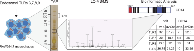
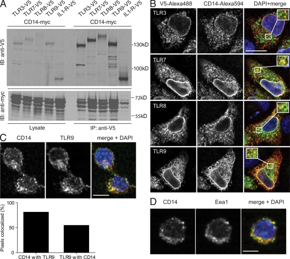
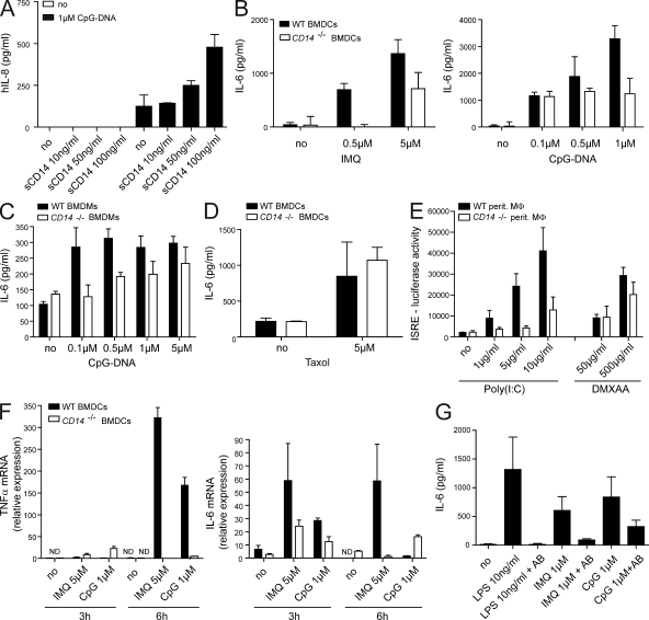
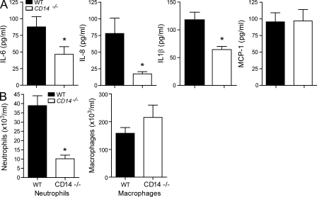
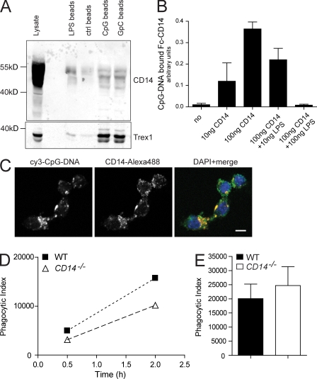
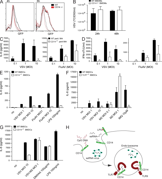
Recognition of pathogens by the innate immune system requires proteins that detect conserved molecular patterns. Nucleic acids are recognized by cytoplasmic sensors as well as by endosomal Toll-like receptors (TLRs). It has become evident that TLRs require additional proteins to be activated by their respective ligands. In this study, we show that CD14 (cluster of differentiation 14) constitutively interacts with the MyD88-dependent TLR7 and TLR9. CD14 was necessary for TLR7- and TLR9-dependent induction of proinflammatory cytokines in vitro and for TLR9-dependent innate immune responses in mice. CD14 associated with TLR9 stimulatory DNA in precipitation experiments and confocal imaging. The absence of CD14 led to reduced nucleic acid uptake in macrophages. Additionally, CD14 played a role in the stimulation of TLRs by viruses. Using various types of vesicular stomatitis virus, we showed that CD14 is dispensable for viral uptake but is required for the triggering of TLR-dependent cytokine responses. These data show that CD14 has a dual role in nucleic acid-mediated TLR activation: it promotes the selective uptake of nucleic acids, and it acts as a coreceptor for endosomal TLR activation.


Figures

Figure 1.
Figure 1.
Schematic illustration of the TLR interaction proteomic approach. Murine endosomal TLR3, TLR7, TLR8, and TLR9 were stably expressed in the macrophage cell line RAW264.7 by retroviral transduction. After TAP of the receptor complexes, eluates were analyzed by one-dimensional SDS-PAGE and silver stained. 20 gel slices were excised from each lane, the proteins were digested, and the peptides were analyzed by LC-MSMS. All bait TLRs were recovered with the average amount of peptides and sequence coverage as indicated in the table. At ∼55 kD, several peptides were identified from CD14. A schematic illustration of CD14 is depicted with the domains shown (signal peptide in red and leucine-rich repeats in blue) and the location of the identified peptides (black bars). For each TLR, the average number of peptides (av p.) identified and the sequence coverage (av%sc) of CD14 are indicated in the table.
Figure 2.
Figure 2.
CD14 is an interactor of TLR3, TLR7, TLR8, and TLR9. (A) Myc-tagged CD14 was double transfected in Hek293T cells with either one V5-tagged endosomal TLR or V5–IL-1R as a negative control. 48 h after transfection, V5-tagged proteins were immunoprecipitated out of cell lysates using V5 agarose. Coprecipitation of CD14 was analyzed by Western blotting for myc and V5. IB, immunoblot. (B) Myc-CD14 was cotransfected with V5-TLR3, -TLR7, -TLR8, or -TLR9 into HeLaS3 cells. Colocalization was analyzed by confocal imaging. Representative areas of overlapping localization are shown magnified in the insets in the merge panel. (C) Images of a confocal section of endogenous CD14 and TLR9 in unstimulated RAW264.7 macrophages. (bottom) Quantification of three-dimensional colocalization analysis. Graph shows the percentage of pixels positive for CD14 colocalizing with pixels positive for TLR9 and the percentage of pixels positive for TLR9 colocalizing with pixels positive for CD14. (D) Colocalization analysis of the endosomal marker protein Eea1 and CD14 in unstimulated RAW264.7 macrophages. Data in A–D are representative of three independent experiments. Quantitation in C is a mean of two independent experiments with each n = 600. Bars: (B) 10 µm; (C and D) 5 µm.
Figure 3.
Figure 3.
CD14 is required for the proinflammatory response to imiquimod and CpG-DNA. (A) Hek293-TLR9 cells were preincubated with the indicated concentrations of sCD14 30 min before stimulation with 1 µM CpG or mock control (no). Culture supernatants were harvested 8 h later, and the levels of human IL-8 were determined. Data are presented as mean ± SD and are representative of two independent experiments, each performed in biological duplicates. (B–D) BMDCs (B and D) or BMDMs (C) from WT or CD14−/− mice were stimulated with CpG-DNA, imiquimod (IMQ), or Taxol for 6 h. IL-6 in supernatants was measured by ELISA. Data are presented as mean ± SD. (E) Peritoneal macrophages were stimulated with Poly(I:C) or DMXAA for 16 h, and type I IFN levels were determined using the IFN luciferase reporter cell line LL171. (F) BMDCs were stimulated for 3 and 6 h as indicated. TRIZOL-extracted RNA was reverse transcribed and used for quantitative real-time PCR to analyze the abundance of transcripts encoding TNF and IL-6. (G) BMDMs from WT mice were incubated with a CD14-specific inhibitory antibody (AB) where indicated 30 min before stimulation with LPS, imiquimod, or CpG-DNA. After 12 h of stimulation, culture supernatants were harvested, and IL-6 protein was measured by ELISA. Data are presented as mean ± SD and are representative of at least three independent experiments, each performed in biological triplicates.
Figure 4.
Figure 4.
CD14 is important for the proinflammatory response to CpG-DNA in vivo. WT or CD14−/− mice were i.p. injected with 8 nmol CpG-DNA. After 4 h, mice were sacrificed. (A) The levels of the proinflammatory cytokines IL-6, KC (IL-8), IL-1β, and MCP-1 in peritoneal lavage were analyzed by ELISA. (B) Peritoneal lavage was taken, and total cells, neutrophils, and macrophages were counted. Differences in cytokine values of WT and CD14−/− stimulation pairs were tested for significance using the one-way analysis of variance and Tukey’s post test. Significant alterations in pattern are marked with an asterisk (*, P < 0.05 vs. WT mice; Mann–Whitney T test). In A, P = 0.0434, P = 0.0005, and P = 0.0029 for IL-6, IL-8, and IL-1β, respectively. In B, P = 0.0002 for neutrophils. Data are presented as mean ± SD and are representative of two independent experiments, each performed with eight mice/group.
Figure 5.
Figure 5.
Association of CD14 with DNA is required for DNA internalization and IL-6 induction. (A) Lysates from untreated RAW264.7 macrophages were incubated for 2 h with streptavidin beads that were coated with either biotinylated LPS or biotinylated CpG-DNA or with GpC-DNA. After four wash steps, bead-bound CD14 or a DNA-binding protein control (Trex1) was visualized by Western blotting. (B) For ELISA-based assay, CpG-DNA–coated ELISA plates were incubated with a soluble CD14-Fc chimera and LPS as competitor. Absorbance of anti–mouse horseradish peroxidase antibody indicates DNA-bound CD14. Data are mean ± SD. (C) 1 µM cy3-CpG-DNA was added to the culture medium of RAW264.7 macrophages. CD14 colocalization with endocytosed fluorescent DNA was visualized by immunostaining and analyzed by confocal imaging. Bar, 5 µm. (D) 1 µM cy3-CpG-DNA was added to the supernatant of WT- or CD14−/−-derived peritoneal macrophages for the indicated time periods (x axis). The uptake of fluorescent DNA by F4/80-positive macrophages was measured by FACS analysis and depicted as phagocytic index (y axis). (E) The uptake of heat-killed E. coli by thioglycollate-elicited peritoneal macrophages of WT and CD14−/− mice was tested and depicted as phagocytic index. Data in E are presented as mean ± SD. All experiments in A–E are representative of at least two independent experiments.
Figure 6.
Figure 6.
CD14 is dispensable for virus uptake but required for the induction of cytokines in ssRNA virus-infected macrophages. (A) Peritoneal macrophages from WT or CD14−/− mice were infected with VSV that expresses a GFP-fused glycoprotein (VSV-GFP; MOI: 1) and analyzed by FACS. The histogram shows GFP positivity of uninfected cells (dashed lines) or cells that were infected for either 4 or 6 h (solid lines). Cells were gated on forward and sideward scatter. (B) Virus accumulation in supernatant of BMDMs that were infected with VSV (MOI: 1) for the indicated time periods. The graph shows the mean virus titer from three independent experiments. Data are mean ± SD. (C–E) Accumulation of IL-6 or type I IFN 14 h after infection of WT and CD14-deficient peritoneal macrophages (C and D) or BMDCs (E) with the indicated MOI of VSV and FluAV or stimulated with LPS. (F and G) BMDCs from WT or TLR7-deficient (F) or WT and CD14-deficient (G) mice were infected with the indicated amounts of VSV or VSV-M2, a mutant which is mainly recognized in the cytoplasm, or stimulated with imiquimod, LPS, or DMXAA. IL-6 accumulation was tested 14 h after stimulation. Data in C–G are presented as mean ± SD and are representative of at least two independent experiments. (H) The model shows that CD14 associates with DNA/ssRNA at the plasma membrane and promotes their endocytosis. ssRNA viruses enter the endosome in a CD14-independent way. Acidification destroys the viral envelope, releasing the viral ssRNA genome. In the endosome, CD14 functions as a coreceptor for TLR7 and TLR9 in the recognition of ssRNA and DNA.

References

    1. Akashi-Takamura S., Miyake K. 2008. TLR accessory molecules. Curr. Opin. Immunol. 20:420–425 10.1016/j.coi.2008.07.001 - DOI - PubMed
    1. Andrejeva J., Childs K.S., Young D.F., Carlos T.S., Stock N., Goodbourn S., Randall R.E. 2004. The V proteins of paramyxoviruses bind the IFN-inducible RNA helicase, mda-5, and inhibit its activation of the IFN-beta promoter. Proc. Natl. Acad. Sci. USA. 101:17264–17269 10.1073/pnas.0407639101 - DOI - PMC - PubMed
    1. Blasius A.L., Beutler B. 2010. Intracellular toll-like receptors. Immunity. 32:305–315 10.1016/j.immuni.2010.03.012 - DOI - PubMed
    1. Bouwmeester T., Bauch A., Ruffner H., Angrand P.O., Bergamini G., Croughton K., Cruciat C., Eberhard D., Gagneur J., Ghidelli S., et al. 2004. A physical and functional map of the human TNF-alpha/NF-kappa B signal transduction pathway. Nat. Cell Biol. 6:97–105 10.1038/ncb1086 - DOI - PubMed
    1. Brinkmann M.M., Spooner E., Hoebe K., Beutler B., Ploegh H.L., Kim Y.M. 2007. The interaction between the ER membrane protein UNC93B and TLR3, 7, and 9 is crucial for TLR signaling. J. Cell Biol. 177:265–275 10.1083/jcb.200612056 - DOI - PMC - PubMed

Publication types

MeSH terms