. 2010 Dec 7;107(49):21116-21.
doi: 10.1073/pnas.1013405107. Epub 2010 Nov 15.

Specific posttranslational modification regulates early events in mammary carcinoma formation

Affiliations

Specific posttranslational modification regulates early events in mammary carcinoma formation

Hua-Bei Guo et al. Proc Natl Acad Sci U S A. .

Abstract

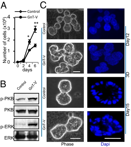
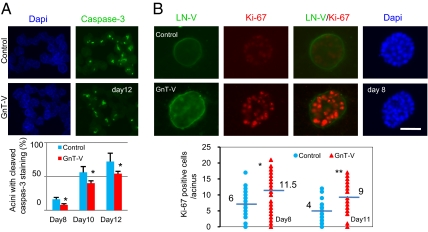
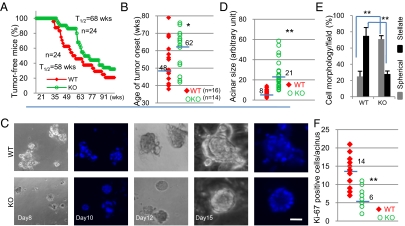
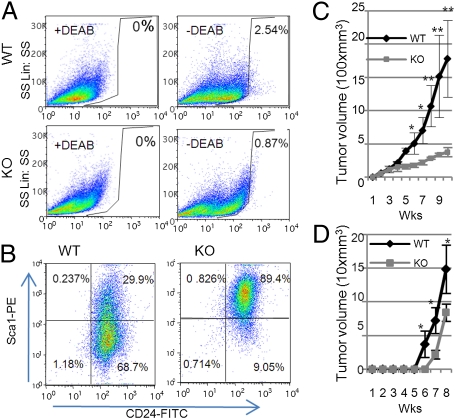
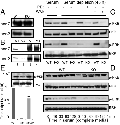
The expression of an enzyme, GnT-V, that catalyzes a specific posttranslational modification of a family of glycoproteins, namely a branched N-glycan, is transcriptionally up-regulated during breast carcinoma oncogenesis. To determine the molecular basis of how early events in breast carcinoma formation are regulated by GnT-V, we studied both the early stages of mammary tumor formation by using 3D cell culture and a her-2 transgenic mouse mammary tumor model. Overexpression of GnT-V in MCF-10A mammary epithelial cells in 3D culture disrupted acinar morphogenesis with impaired hollow lumen formation, an early characteristic of mammary neoplastic transformation. The disrupted acinar morphogenesis of mammary tumor cells in 3D culture caused by her-2 expression was reversed in tumors that lacked GnT-V expression. Moreover, her-2-induced mammary tumor onset was significantly delayed in the GnT-V null tumors, evidence that the lack of the posttranslational modification catalyzed by GnT-V attenuated tumor formation. Inhibited activation of both PKB and ERK signaling pathways was observed in GnT-V null tumor cells. The proportion of tumor-initiating cells (TICs) in the mammary tumors from GnT-V null mice was significantly reduced compared with controls, and GnT-V null TICs displayed a reduced ability to form secondary tumors in NOD/SCID mice. These results demonstrate that GnT-V expression and its branched glycan products effectively modulate her-2-mediated signaling pathways that, in turn, regulate the relative proportion of tumor initiating cells and the latency of her-2-driven tumor onset.


Conflict of interest statement

The authors declare no conflict of interest.

Figures

Fig. 1.
Fig. 1.
Increased expression of GnT-V stimulated cell proliferation and disrupted mammary acinar morphogenesis of MCF-10A cells in 3D culture. (A) Cells were grown on six-well plates and counted at each indicated time point and expressed as the mean ± SD of triplicate samples. *P < 0.05; **P < 0.001. (B) Cells were collected for detection of indicated proteins by immunoblot. (C) Cells were grown in 3D culture for 12 or 15 d. Nuclei (DNA) were stained with DAPI (4′,6-diamidino-2-phenylindole) and visualized by confocal microscopy. Images are representative of three independent experiments. (Scale bars: 25 μm.)
Fig. 2.
Fig. 2.
GnT-V overexpression increased proliferation and reduced sensitivity to anoikis during acinar morphogenesis of MCF-10A cells. (A) Cells were grown in 3D culture for the indicated times and stained for cleaved caspase-3. Graph shows percentages of acini with cleaved caspase-3 staining, which were calculated by counting the number of acini with or without cleaved caspase-3 staining in five different fields and expressed as the mean ± SD. (B) Cells were grown in 3D culture for 8 d and stained for LN-V (green), Ki-67 (red), and DAPI (blue). Graph shows number of Ki-67 positive cells per acinus on day 8 and day 11; 50–100 acini were scored in each group. -, median value; *P < 0.05; **P < 0.001. (Scale bar: 25 μm.)
Fig. 3.
Fig. 3.
Tumor onset was inhibited and the disrupted acinar formation was reversed in her-2/GnT-V KO cells. (A) Kaplan–Meier curves were used to display the latency of her-2–induced mammary tumors in GnT-V WT and KO mice (P < 0.05). T1/2 represents the time point at which 50% of mice in that group developed tumors. (B) Median number of weeks for tumor onset for an individual mouse was calculated from each group of mice under 78 wk of age. (C) Cells were grown in 3D culture for indicated days and stained with DAPI. (DF) Cells were grown in 3D culture for 10 d, individual acinar size was measured (D), cell morphology observed in five different fields was scored (E), and the Ki-67 positive cells per acinus were counted (F). Fifty to 100 acini were scored in each group. -, median value; *P < 0.05; **P < 0.001. (Scale bar: 25 μm.)
Fig. 4.
Fig. 4.
Her-2–induced GnT-V KO tumors displayed a reduced population of TICs and decreased secondary tumor formation in NOD/SCID mice. (A) Aldefluor assays were performed by using tumor cells isolated from primary tumors, and the percentage of Aldefluor-positive cells was determined under similar gating criteria. Data are representative of two independent experiments. (B) Lin epithelial cells purified from either WT or KO tumor cells were analyzed by flow cytometry with labeled Sca1 and CD24 antibodies to identify Sca1/CD24+ populations. Data are representative of two independent experiments. (C) Primary tumor cells (1 × 10⁶) were injected into No. 4 mammary fat pads of NOD/SCID mice (n = 5), and secondary tumor growth was observed up to 10 wk. Tumor volume was measured and expressed as the mean ± SD. (D) Aldefluor-positive cells (103) were sorted from secondary tumor tissues formed by implantation of either WT or KO cells and were then injected into No. 4 mammary fat pads of NOD/SCID mice (n = 3). Tumor formation was observed for 8 wk. *P < 0.05; **P < 0.005.
Fig. 5.
Fig. 5.
Deletion of GnT-V inhibited her-2–mediated downstream signaling pathways. (A) Cell surface proteins were labeled with NHS-LC-biotin, followed by streptavidin immunoprecipitation (IP). Precipitated proteins were subjected to detection of indicated proteins by immunoblot. (B) Cells were lysed for L-PHA IP, and both precipitated L-PHA bound proteins (lane 1 and 2) and cell lysates (used as control, lane 3 and 4) were subjected to detection of indicated proteins by immunoblot. (C) Cells grown either in serum-containing or serum-free media for 2 d with or without PD98059 (PD, 20 μM) or wortmannin (WM, 200 nM) were collected for detection of indicated proteins by immunoblot. (D) Cells grown in serum-free media for 2 d were stimulated with complete growth media for indicated times and collected for detection of indicated proteins by immunoblot. (E) GnT-V KO cells transiently transfected with GnT-V cDNA (KO/V+) were collected for determination of GnT-V transcripts by qRT-PCR (Lower) and immunoblot for detection of indicated proteins (Upper).

Similar articles

Cited by

References

    1. Carlsson J, et al. HER2 expression in breast cancer primary tumours and corresponding metastases. Original data and literature review. Br J Cancer. 2004;90:2344–2348. - PMC - PubMed
    1. Ménard S, et al. HER2 overexpression in various tumor types, focussing on its relationship to the development of invasive breast cancer. Ann Oncol. 2001;12(Suppl 1):S15–S19. - PubMed
    1. Guy CT, et al. Expression of the neu protooncogene in the mammary epithelium of transgenic mice induces metastatic disease. Proc Natl Acad Sci USA. 1992;89:10578–10582. - PMC - PubMed
    1. Sorkin A, Goh LK. Endocytosis and intracellular trafficking of ErbBs. Exp Cell Res. 2009;315:683–696. - PubMed
    1. Holbro T, Civenni G, Hynes NE. The ErbB receptors and their role in cancer progression. Exp Cell Res. 2003;284:99–110. - PubMed

Publication types

MeSH terms

Substances

LinkOut - more resources