. 2010 Dec 7;107(49):21170-5.
doi: 10.1073/pnas.1011949107. Epub 2010 Nov 15.

Stable and dynamic cortical electrophysiology of induction and emergence with propofol anesthesia

Affiliations

Stable and dynamic cortical electrophysiology of induction and emergence with propofol anesthesia

Jonathan D Breshears et al. Proc Natl Acad Sci U S A. .

Abstract

The mechanism(s) by which anesthetics reversibly suppress consciousness are incompletely understood. Previous functional imaging studies demonstrated dynamic changes in thalamic and cortical metabolic activity, as well as the maintained presence of metabolically defined functional networks despite the loss of consciousness. However, the invasive electrophysiology associated with these observations has yet to be studied. By recording electrical activity directly from the cortical surface, electrocorticography (ECoG) provides a powerful method to integrate spatial, temporal, and spectral features of cortical electrophysiology not possible with noninvasive approaches. In this study, we report a unique comprehensive recording of invasive human cortical physiology during both induction and emergence from propofol anesthesia. Propofol-induced transitions in and out of consciousness (defined here as responsiveness) were characterized by maintained large-scale functional networks defined by correlated fluctuations of the slow cortical potential (<0.5 Hz) over the somatomotor cortex, present even in the deeply anesthetized state of burst suppression. Similarly, phase-power coupling between θ- and γ-range frequencies persisted throughout the induction and emergence from anesthesia. Superimposed on this preserved functional architecture were alterations in frequency band power, variance, covariance, and phase-power interactions that were distinct to different frequency ranges and occurred in separable phases. These data support that dynamic alterations in cortical and thalamocortical circuit activity occur in the context of a larger stable architecture that is maintained despite anesthetic-induced alterations in consciousness.


Conflict of interest statement

The authors declare no conflict of interest.

Figures

Fig. 1.
Fig. 1.
The SCP somatomotor network is maintained throughout induction and recovery. The correlation of the SCP at all electrodes with a seed electrode on motor cortex is shown in five contiguous nonoverlapping epochs for (A) induction and (B) recovery. Correlation coefficients are plotted at their respective electrode locations on a standardized Montreal Neurological Institute brain. Distributions generated by epoch subsampling were not statistically different (P < 0.05). Subject 4 was excluded because of large seizure foci in the motor cortex; subject 8 was excluded because the grid did not cover the motor cortex.
Fig. 2.
Fig. 2.
The SCP somatomotor network is maintained during burst suppression. (A) The correlation coefficient of the SCP with a motor cortex seed electrode in three subjects during the period of burst suppression shown (blue trace). Mean correlation structure is shown during (B) burst and (C) suppression intervals only.
Fig. 3.
Fig. 3.
Topographic absolute power changes during induction and recovery. The robust fit coefficient, or slope (P < 0.001), of the absolute band power trend at each electrode during induction (A) and recovery (B) is shown for the nine frequency bands. The mean and 95% confidence interval across all subjects’ electrodes is shown at the bottom. Total electrode coverage is shown in Insets. Blue, left hemispheric coverage. Yellow, right hemispheric coverage. Discrepancy in left hemispheric coverage between induction and emergence reflects absence of emergence data from subjects 7 and 8 and absence of induction data from subject 6.
Fig. 4.
Fig. 4.
Trends in μ, β, and γ1 power variability and covariance between distant cortical sites. (A) Average normalized μ, β, and γ1 power from an exemplar electrode demonstrating increased variance during induction that decreases on recovery. (B) The mean intraband power correlation between electrodes. Power in the μ, β1–2, and γ1 bands shows increasing correlation during induction and a decrease during recovery.
Fig. 5.
Fig. 5.
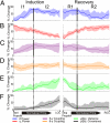
(A–F) Phases of change in spectral power, variance, covariance, and phase–power coupling during induction and recovery. Solid vertical lines demarcate mean knot sites, where segments were found to be separable with adaptive piecewise regression. These demarcations were used to separate the different phases of inductions and recovery. All trends reflect the smoothed median and bootstrapped 95% confidence interval. I1, induction phase 1; I2, induction phase 2; R1, recovery phase 1; R2, recovery phase 2.
Fig. 6.
Fig. 6.
Phase–power correlations during induction and recovery. (A) The mean correlation coefficient across all subjects (P < 0.01, Bonferroni corrected) between the signal phase (<25 Hz) and the power in the nine frequency bands. (B) Data from an exemplar electrode illustrating the relationship between 1- to 4-Hz phase and high-frequency power that emerges during induction and dissipates on emergence. The average γ2 power fluctuations (black) are increasingly and decreasingly correlated to the phase of the 1- to 4-Hz signal component (blue) on induction and recovery, respectively. High-frequency oscillations (red) illustrate this amplitude modulation.

References

    1. Franks NP. General anaesthesia: From molecular targets to neuronal pathways of sleep and arousal. Nat Rev Neurosci. 2008;9:370–386. - PubMed
    1. Alkire MT, Haier RJ, Fallon JH. Toward a unified theory of narcosis: Brain imaging evidence for a thalamocortical switch as the neurophysiologic basis of anesthetic-induced unconsciousness. Conscious Cogn. 2000;9:370–386. - PubMed
    1. Fiset P, et al. Brain mechanisms of propofol-induced loss of consciousness in humans: A positron emission tomographic study. J Neurosci. 1999;19:5506–5513. - PMC - PubMed
    1. White NS, Alkire MT. Impaired thalamocortical connectivity in humans during general-anesthetic-induced unconsciousness. Neuroimage. 2003;19:402–411. - PubMed
    1. Ferrarelli F, et al. Breakdown in cortical effective connectivity during midazolam-induced loss of consciousness. Proc Natl Acad Sci USA. 2010;107:2681–2686. - PMC - PubMed

Publication types

MeSH terms

LinkOut - more resources