. 2010 Dec 7;107(49):21040-5.
doi: 10.1073/pnas.1012248107. Epub 2010 Nov 15.

Neural crest origin of olfactory ensheathing glia

Affiliations

Neural crest origin of olfactory ensheathing glia

Perrine Barraud et al. Proc Natl Acad Sci U S A. .

Abstract

Olfactory ensheathing cells (OECs) are a unique class of glial cells with exceptional translational potential because of their ability to support axon regeneration in the central nervous system. Although OECs are similar in many ways to immature and nonmyelinating Schwann cells, and can myelinate large-diameter axons indistinguishably from myelination by Schwann cells, current dogma holds that OECs arise from the olfactory epithelium. Here, using fate-mapping techniques in chicken embryos and genetic lineage tracing in mice, we show that OECs in fact originate from the neural crest and hence share a common developmental heritage with Schwann cells. This explains the similarities between OECs and Schwann cells and overturns the existing dogma on the developmental origin of OECs. Because neural crest stem cells persist in adult tissue, including skin and hair follicles, our results also raise the possibility that patient-derived neural crest stem cells could in the future provide an abundant and accessible source of autologous OECs for cell transplantation therapy for the injured central nervous system.


Conflict of interest statement

The authors declare no conflict of interest.

Figures
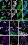
Fig. 1.
Fig. 1.
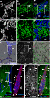
The olfactory placode does not form OECs. Unilateral isotopic grafts of GFPchick ANF at the 3–5 ss label the olfactory epithelium (and prospective olfactory bulb) on the operated side at E4.5 (A and C) and E10.25 (E and F, which show examples from two different embryos). At E4.5, all of the olfactory placode-derived cells on the olfactory nerve seem to be neurons, expressing the neuronal markers neuronal β-III tubulin (B–B2) and/or HuC/D (D–D2). At E10.25, p75NTR-positive OEC processes (red) ensheath bundles of olfactory axons (blue), both in the lamina propria beneath the olfactory epithelium (G and G1) and more proximally along the nerve (H–H2). Virtually none of the p75NTR-positive OEC processes (red) are graft-derived (G1, H1, and H2). Arrowheads in G and G1 indicate examples of p75NTR-positive/GFP-negative OEC processes ensheathing bundles of GFP-positive olfactory axons. Occasional p75NTR-positive/GFP-positive processes (arrow in HH2) are most likely derived from the few NCCs contributed by the graft (scattered GFP-positive cells are seen in the nasal septum and frontonasal mesenchyme: arrowheads in HH2). nβ3-tub, neuronal β-III tubulin; OB, prospective olfactory bulb; OE, olfactory epithelium; ON, olfactory nerve; ns, nasal septum.
Fig. 2.
Fig. 2.
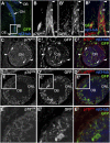
Avian neural crest cells form OECs on the olfactory nerve. Isotopic grafts of GFPchick (A–B2 and EF2) or quail (CD) midbrain-level neural fold at the 5-ss label migratory NCCs that colonize the olfactory nerve as well as the surrounding frontonasal mesenchyme. At E4.5 (AB2), nonneuronal NCCs are associated with the olfactory nerve. (Faint green staining in the apical olfactory epithelium is background.) At E5.5 (CD), many Sox10-positive cells are seen on the olfactory nerve on the grafted side of the embryo. Quail NCC-derived cells (green nuclei) on the olfactory nerve express Sox10 (D). At E6.5 (EF2), a significant proportion of olfactory nerve-associated GFPchick NCCs and their processes express the OEC marker P0 (arrowheads, F–F2) (22). nβ3-tub, neuronal β-III tubulin; OB, olfactory bulb; OE, olfactory epithelium; ON, olfactory nerve.
Fig. 3.
Fig. 3.
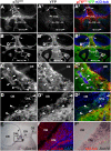
Chick neural crest cells form OECs along the length of the olfactory nerve from the lamina propria to the olfactory bulb. Isotopic grafts of GFPchick midbrain-level neural fold at the 5 ss label migratory NCCs. At E10.25, p75NTR-positive OECs are associated with p75NTR-positive olfactory axons/neurons in the lamina propria (A and B). These p75NTR-positive OECs, like the rest of the lamina propria and the cribriform plate, are GFPchick NCC-derived (arrowheads highlight examples) (A1, A2, B1, and B2). Similarly, the p75NTR-positive OEC processes ensheathing olfactory axon bundles in the olfactory nerve (C and D) are from GFPchick NCC-derived cells (C1, C2, D1, and D2), whose cell bodies can be seen as brighter patches of immunofluorescence between the axon bundles (the p75NTR immunoreactivity is confined to their cell processes). P0-positive OEC processes (E) are also from GFPchick NCC-derived cells (E1 and E2). (F) Low-power composite view showing that GFPchick NCC-derived cells not only surround the olfactory bulb but also are found throughout the ONL. [The scattered GFPchick NCC-derived cells in the olfactory bulb are smooth muscle actin-positive pericytes associated with the forebrain vasculature (also see Fig. S3).] (GG2) Higher-power view showing GFPchick NCC-derived cells throughout the ONL, with cells in the outer layer expressing both p75NTR (blue) and P0 (red), and cells in the inner layer expressing P0 but not p75NTR. (H–H2) In a different E10.25 embryo, the olfactory bulb is surrounded by GFPchick NCC-derived cells, and almost all of the P0-positive OECs inside the olfactory bulb are GFPchick NCC-derived (arrowheads highlight examples). Arrows highlight some P0-positive OECs that are not GFPchick graft-derived; these are likely to be derived from host NCCs. CP, cribriform plate; LP, lamina propria; nβ3-tub, neuronal β-III tubulin; OE, olfactory epithelium; ON, olfactory nerve; ONL, olfactory nerve layer.
Fig. 4.
Fig. 4.
Neural crest cells from the hindbrain can form OECs. (A–B2) At E6.5, GFPchick NCC-derived cells, at least some of which express p75NTR (arrowheads highlight examples), are present on the olfactory nerve after heterotopic grafts of rhombomeres 4–6 to the rostral midbrain. (C–C2) At E9.5–10.25, GFPchick hindbrain NCC-derived cells, at least some of which express p75NTR (arrowheads highlight examples), ensheath bundles of axons in the olfactory nerve. (D–E2) At E9.5–10.25, p75NTR-positive GFPchick hindbrain NCC-derived cells are abundantly found in the ONL. nβ3-tub, neuronal β-III tubulin; OB, olfactory bulb; OE, olfactory epithelium; ON, olfactory nerve; ONL, olfactory nerve layer.
Fig. 5.
Fig. 5.
Genetic lineage-tracing using Wnt1Cre;R26RYFP embryos shows that mouse neural crest cells form OECs. (A–A2) No above-background YFP immunoreactivity is seen in the olfactory region of an E13.5 Wnt1Cre-negative embryo from a cross between Wnt1Cre and R26RYFP reporter mice (compare A1 and A2 with B1 and B2). (B–B2) In a Wnt1Cre;R26RYFP littermate, YFP-positive NCC-derived cells are found throughout the frontonasal mass, as well as in the ONL. The olfactory epithelia and vomeronasal organ epithelia show no above-background YFP immunoreactivity (compare B1 and B2 with A1 and A2). (C–C2) p75NTR-positive OECs associated with olfactory axons/neurons in the lamina propria are NCC-derived (YFP-positive), as is the rest of the lamina propria. Arrowheads highlight examples. (D–D2) YFP-positive NCC-derived cells, some of which express p75NTR, are found throughout the ONL. Arrowheads highlight examples of p75NTR-positive OECs in the ONL. (E–F2) In situ hybridization on sections of a wild-type E17.5 embryo shows that Sox10 is a marker for mouse OECs. [Sox10 expression in the olfactory epithelium likely represents developing Bowman's glands (Fig. S8).] LP, lamina propria; nβ3-tub, neuronal β-III tubulin; OB, olfactory bulb; OE, olfactory epithelium; ON, olfactory nerve; ONL, olfactory nerve layer; vno, vomeronasal organ.

References

    1. Barnett SC, Riddell JS. Olfactory ensheathing cell transplantation as a strategy for spinal cord repair: What can it achieve? Nat Clin Pract Neurol. 2007;3:152–161. - PubMed
    1. Raisman G, Li Y. Repair of neural pathways by olfactory ensheathing cells. Nat Rev Neurosci. 2007;8:312–319. - PubMed
    1. Richter MW, Roskams AJ. Olfactory ensheathing cell transplantation following spinal cord injury: Hype or hope? Exp Neurol. 2008;209:353–367. - PubMed
    1. Kawaja MD, Boyd JG, Smithson LJ, Jahed A, Doucette R. Technical strategies to isolate olfactory ensheathing cells for intraspinal implantation. J Neurotrauma. 2009;26:155–177. - PubMed
    1. Couly GF, Le Douarin NM. Mapping of the early neural primordium in quail-chick chimeras. I. Developmental relationships between placodes, facial ectoderm, and prosencephalon. Dev Biol. 1985;110:422–439. - PubMed

Publication types