Review
. 2010 Dec;62(4):726-59.
doi: 10.1124/pr.110.002733.

Neutrophil elastase, proteinase 3, and cathepsin G as therapeutic targets in human diseases

Affiliations
Review

Neutrophil elastase, proteinase 3, and cathepsin G as therapeutic targets in human diseases

Brice Korkmaz et al. Pharmacol Rev. 2010 Dec.

Abstract

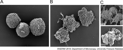
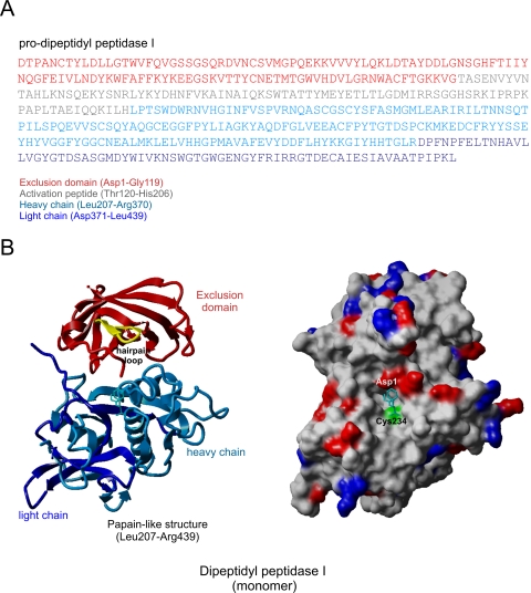
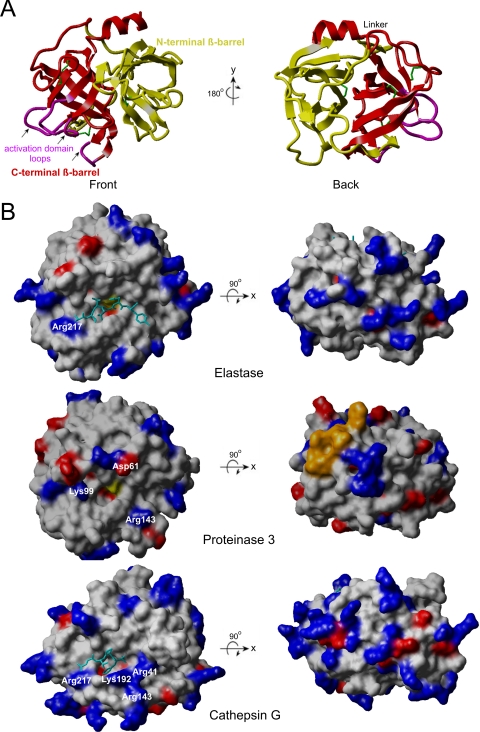
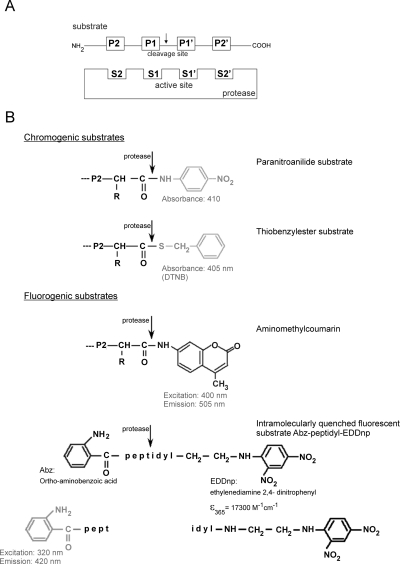
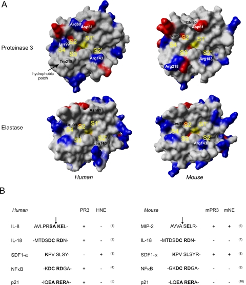
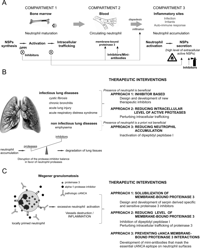
Polymorphonuclear neutrophils are the first cells recruited to inflammatory sites and form the earliest line of defense against invading microorganisms. Neutrophil elastase, proteinase 3, and cathepsin G are three hematopoietic serine proteases stored in large quantities in neutrophil cytoplasmic azurophilic granules. They act in combination with reactive oxygen species to help degrade engulfed microorganisms inside phagolysosomes. These proteases are also externalized in an active form during neutrophil activation at inflammatory sites, thus contributing to the regulation of inflammatory and immune responses. As multifunctional proteases, they also play a regulatory role in noninfectious inflammatory diseases. Mutations in the ELA2/ELANE gene, encoding neutrophil elastase, are the cause of human congenital neutropenia. Neutrophil membrane-bound proteinase 3 serves as an autoantigen in Wegener granulomatosis, a systemic autoimmune vasculitis. All three proteases are affected by mutations of the gene (CTSC) encoding dipeptidyl peptidase I, a protease required for activation of their proform before storage in cytoplasmic granules. Mutations of CTSC cause Papillon-Lefèvre syndrome. Because of their roles in host defense and disease, elastase, proteinase 3, and cathepsin G are of interest as potential therapeutic targets. In this review, we describe the physicochemical functions of these proteases, toward a goal of better delineating their role in human diseases and identifying new therapeutic strategies based on the modulation of their bioavailability and activity. We also describe how nonhuman primate experimental models could assist with testing the efficacy of proposed therapeutic strategies.


Figures

Fig. 1.
Fig. 1.
Polymorphonuclear neutrophil. Quiescent (A) and chemically activated (B) neutrophils purified from peripheral blood. C, PMA-activated neutrophils embedded within NET and neutrophil spreading on insoluble elastin.
Fig. 2.
Fig. 2.
Structure of human pro-dipeptidyl peptidase I. A, amino acid sequence of human pro-dipeptidyl peptidase I. Primary sequences corresponding to exclusion domain (Asp1–Gly119), activation peptide (Thr120–His206), heavy chain (Leu207–Arg370), and light chain (Asp371–Leu439) are colored in red, gray, light, and dark blue, respectively. B, ribbon representation and solvent-accessible surfaces of the DPPI functional monomer in complex with the inhibitor Gly-Phe-CHN2 [Protein Data Bank 1K3B (Turk et al., 2001), 2DJF, and 2DJG (Mølgaard et al., 2007)]. Color codes in the ribbon plot are the same as in A except that Asp1 in the hairpin loop is colored yellow (left). Solvent-accessible surfaces with positive or negative electrostatic potential are colored dark blue and red, respectively (right). The side chain of the catalytic Cys234 is showed with a cyan stick. The figures have been created with Yasara (http://www.yasara.org).
Fig. 3.
Fig. 3.
Three-dimensional architecture of the trypsin/chymotrypsin family and structural differences between neutrophil elastase, proteinase 3, and cathepsin G. A, ribbon plot of neutrophil elastase showing the characteristics of the trypsin/chymotrypsin family, the two asymmetric β-barrels and the C-terminal α-helix, front view on the left, back view after a rotation of 180° around a vertical y-axis on the right. N- and C-terminal β-barrels are presented in red and yellow, respectively. The three flexible loops of the C-terminal β-barrels forming the activation domain are colored in pink and indicated by arrows. Disulfide bounds are depicted in green. B, the solvent accessible surface based on the atom coordinates of elastase [Protein Data Bank 1PPF (Bode et al., 1986)], proteinase 3 [Protein Data Bank 1FUJ (Fujinaga et al., 1996)], and cathepsin G [1CGH (Hof et al., 1996)] is colored to show its positive (blue) and negative (red) electrostatic potential. Surface-accessible residues forming a hydrophobic patch on proteinase 3 are depicted in orange. The loop of the inhibitor turkey ovomucoid third domain (P4–P3′) complexed to elastase and the irreversible phosphonate inhibitor Suc-Val-Pro-PheP-(OPh)2 complexed to cathepsin G are given as a cyan stick model. The serine of the catalytic triad is yellow. Ribbon plot of neutrophil elastase and the molecular surfaces are generated with Yasara (http://www.yasara.org).
Fig. 4.
Fig. 4.
Synthetic chromogenic and fluorogenic substrates of NSP. A, schematic diagram illustrating the Schechter and Berger (1967) nomenclature. This nomenclature defines a linear topology of the interactions between a protease and a substrate: S subsites on the protease accommodate P residues of the substrate upstream of the scissile bond, whereas S′ subsites accommodate P′ residues from the C-terminal part of the substrate, downstream of the cleavage site. Numbering starts with the first residue next to the cleavage site, P1 and P1′, and continues toward the N terminus (P2, P3, P4, etc.) and the C terminus (P2′, P3′, P4′, etc.). B, chromogenic substrates commonly used to measure the activity of NSPs, peptidyl-paranitroanilides (top), peptidyl-thiobenzylesters with chromogenic groups in gray. Paranitroanilide may be detected by direct measurement at 410 nm, but a coupled assay with a thiodisulfide reagent such as DTNP is required for measuring the absorbance of the released thiobenzyl group. Fluorogenic aminomethylcoumarin substrates contain a fluorescent group that is released by proteolytic cleavage. FRET substrates contain a fluorescent and a quenching group as a donor/acceptor pair [here a fluorescent ortho-aminobenzoyl (Abz) group and a N-(2,4-dinitrophenyl ethylenediamine (EDDnp) quenching group] at their N- and C-terminal ends, respectively. Fluorescence is released after cleavage of any peptide bond within the amino acid sequence (shown in gray). FRET substrates are convenient tools to investigate the protease specificity around the cleavage site. DTNB, 5,5′-dithiobis(2-nitrobenzoic acid).
Fig. 5.
Fig. 5.
A, interspecies comparisons of the active site region of proteinase 3 and neutrophil elastase from men and mice. Values of electrostatic potential were mapped onto the solvent accessible surface of each protease with positively and negatively charged residues colored in blue and red, respectively. Positions of the subsites S4 to S2′ are shown in yellow. Key residues that lie in close vicinity to the substrate binding pockets are labeled by their three-letter codes. The hydrophobic patch in human PR3 is missing in the mouse homolog as the Trp218 residue is substituted by an Arg. B, sequences derived from natural human and mouse substrates that are cleaved by human and murine NE and PR3. Arrows indicate identified and potential cleavage sites. IL-8: (Padrines et al., 1994) (1); IL-18: putative cleavage sites [(2) and (7)]; SDF-1α: (Valenzuela-Fernandez et al., 2002) (3), putative cleavage site (8); NFκB: (Preston et al., 2002) (4), NFκB (Kalupov et al., 2009) (9); p21 (Dublet et al., 2005) (5), T. Kalupov et al., unpublished data (10); and MIP-2 (T. Dau and D. E. Jenne, unpublished data) (6). The structural models of murine proteases were derived from X-ray structures of human homologs by homology modeling (http://www.expasy.ch).
Fig. 6.
Fig. 6.
A, interventional options for the control of neutrophil proteases. The different compartments in which the exposure to NSPs can be reduced and the biological impact of this intervention are shown. B, the different therapeutic approaches to rebalance the altered protease/antiprotease equilibrium in neutrophil-associated inflammatory lung diseases. C, specification for the treatment of Wegener granulomatosis, which is characterized by excessive ANCA-induced activation of locally primed PR3-positive neutrophils and subsequent small vessel destruction.

Similar articles

Cited by

References

    1. Abraham CR, Selkoe DJ, Potter H. (1988) Immunochemical identification of the serine protease inhibitor alpha 1-antichymotrypsin in the brain amyloid deposits of Alzheimer's disease. Cell 52:487–501 - PubMed
    1. Adkison AM, Raptis SZ, Kelley DG, Pham CT. (2002) Dipeptidyl peptidase I activates neutrophil-derived serine proteases and regulates the development of acute experimental arthritis. J Clin Invest 109:363–371 - PMC - PubMed
    1. Akira S, Uematsu S, Takeuchi O. (2006) Pathogen recognition and innate immunity. Cell 124:783–801 - PubMed
    1. Akita K, Ohtsuki T, Nukada Y, Tanimoto T, Namba M, Okura T, Takakura-Yamamoto R, Torigoe K, Gu Y, Su MS, et al. (1997) Involvement of caspase-1 and caspase-3 in the production and processing of mature human interleukin 18 in monocytic THP.1 cells. J Biol Chem 272:26595–26603 - PubMed
    1. Akk AM, Simmons PM, Chan HW, Agapov E, Holtzman MJ, Grayson MH, Pham CT. (2008) Dipeptidyl peptidase I-dependent neutrophil recruitment modulates the inflammatory response to Sendai virus infection. J Immunol 180:3535–3542 - PMC - PubMed

Publication types

MeSH terms