Review
. 2010 Dec;62(4):760-81.
doi: 10.1124/pr.110.003723.

Drug-induced long QT syndrome

Affiliations
Review

Drug-induced long QT syndrome

Prince Kannankeril et al. Pharmacol Rev. 2010 Dec.

Abstract

The drug-induced long QT syndrome is a distinct clinical entity that has evolved from an electrophysiologic curiosity to a centerpiece in drug regulation and development. This evolution reflects an increasing recognition that a rare adverse drug effect can profoundly upset the balance between benefit and risk that goes into the prescription of a drug by an individual practitioner as well as the approval of a new drug entity by a regulatory agency. This review will outline how defining the central mechanism, block of the cardiac delayed-rectifier potassium current I(Kr), has contributed to defining risk in patients and in populations. Models for studying risk, and understanding the way in which clinical risk factors modulate cardiac repolarization at the molecular level are discussed. Finally, the role of genetic variants in modulating risk is described.


Figures

Fig. 1.
Fig. 1.
Continuous recording from a patient who had recently begun receiving sotalol. During AF, there is irregularity of ventricular response, creating frequent short-long-short cycles but there is minimal change in QT intervals (top). After electrical cardioversion, the QT interval increased dramatically to 0.64 s (middle), and an episode of torsades de pointes is triggered (bottom). [Reprinted from Darbar D, Kimbrough J, Jawaid A, McCray R, Ritchie MD, and Roden DM (2008) Persistent atrial fibrillation is associated with reduced risk of torsades de pointes in patients with drug-induced long QT syndrome. J Am Coll Cardiol 51:836–842. Copyright © 2008 Elsevier, Inc. Used with permission.]
Fig. 2.
Fig. 2.
An example of drug-induced long QT syndrome. A common feature is a pause (often after an ectopic beat) with deranged repolarization in the following cycle. A 12-lead ECG recorded from a 79-year-old patient with advanced heart disease who had recently begun taking dofetilide. The abnormal QT interval is followed by a pause (star) and then four beats of polymorphic ventricular tachycardia (torsades de pointes). Sustained torsades de pointes then occurs after another pause.
Fig. 3.
Fig. 3.
Rhythm recording from a 70-year-old woman with renal dysfunction recently treated with sotalol for AF. She converted to sinus rhythm several minutes before this recording. It shows a typical episode of torsades de pointes with a four-beat run of polymorphic ventricular tachycardia, a pause, and a sinus beat with a long and deformed QT interval (arrow), interrupted by a another episode of TdP. This pattern of onset with a short cycle followed by a long one followed by a short one is typical of drug-induced torsades de pointes. Risk factors in this case included female sex, the administration of sotalol in a patient with renal failure (causing increased drug levels), and recent conversion from AF.
Fig. 4.
Fig. 4.
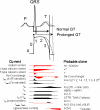
The relationship between ionic currents and the duration of cardiac repolarization recorded from the ECG and the myocardial action potential. Top, idealized ECG recording aligned with a schematized ventricular myocyte action potential. A balance between inward (red) and outward (black) currents controls repolarization, and a relative increase of outward over inward currents drives repolarization to completion. The action potential is initiated by inward sodium current (phase 0) and proceeds through early (phases 1 and 2) and late (phase 3) stages of repolarization. Increases in net inward current prolong repolarization. The “plateau” phases (2 and 3) are vulnerable to minor increases in net inward current, which can initiate early afterdepolarizations that are one likely cause of torsades de pointes LQTS. [Adapted from Anderson ME, Darbar D and Kannankeril P (2007) Genetics of arrhythmias, in Textbook of Cardiovascular Medicine (Topol EJ ed), Lippincott Williams & Wilkins, Philadelphia, PA. Copyright © 2007 Lippincott Williams & Wilkins. Used with permission.]
Fig. 5.
Fig. 5.
Mechanisms of proarrhythmia associated with human ether-à-go-go-related gene (HERG) channel blockade. Blockade of the HERG channel produces prolongation of the QT interval (blue) and generates an EAD (red) in the cardiac action potential. These changes, which are heterogeneous across the ventricular wall, create a substrate for TdP. In this example, torsades de pointes degenerates into ventricular fibrillation. [Adapted from Roden DM and Viswanathan PC (2005) Genetics of acquired long QT syndrome. J Clin Invest 115:2025–2032. Copyright © 2005 American Society for Clinical Investigation. Used with permission.]
Fig. 6.
Fig. 6.
The concept of reduced repolarization reserve. Patient 1 is prescribed a QT-prolonging drug and has minimal or no prolongation of the QT interval. In contrast, patient 2, given the same QT-prolonging drug at the same dosage, develops marked prolongation of the QT interval. This has been termed “reduced repolarization reserve” and as discussed further in section III.D, repolarization is a physiologically redundant process such that IKr block will not result in markedly prolonged repolarization; that is, the system displays some “reserve.” However, otherwise subclinical lesions in other components of the repolarization system, such as reduction of IKs as a result of genetic factors or hypokalemia, may become apparent as marked QT prolongation when IKr is reduced.
Fig. 7.
Fig. 7.
Luo Rudy simulations showing the concept of repolarization reserve. The blue line shows the effect of reducing IKr by 15%, as might be expected in a subtle congenital long QT syndrome mutation. The green line shows the expected prolongation of the control action potential resulting from 75% IKr blockade. The red line shows the effect of the same degree of drug blockade applied to the simulation with 15% IKs blockade. Not only is there marked exaggeration of action potential prolongation, but an EAD with a triggered upstroke is also generated; L-type calcium current generates the upstroke in this model. [Reprinted from Roden DM and Viswanathan PC (2005) Genetics of acquired long QT syndrome. J Clin Invest 115:2025–2032. Copyright © 2005 American Society for Clinical Investigation. Used with permission.]

Similar articles

Cited by

References

    1. Abbott GW, Sesti F, Splawski I, Buck ME, Lehmann MH, Timothy KW, Keating MT, Goldstein SA. (1999) MiRP1 forms IKr potassium channels with HERG and is associated with cardiac arrhythmia. Cell 97:175–187 - PubMed
    1. Abrahamsson C, Palmer M, Ljung B, Duker G, Bäärnhielm C, Carlsson L, Danielsson B. (1994) Induction of rhythm abnormalities in the fetal rat heart. A tentative mechanism for the embryotoxic effect of the class III antiarrhythmic agent almokalant. Cardiovasc Res 28:337–344 - PubMed
    1. Aiba T, Shimizu W, Inagaki M, Noda T, Miyoshi S, Ding WG, Zankov DP, Toyoda F, Matsuura H, Horie M, et al. (2005) Cellular and ionic mechanism for drug-induced long QT syndrome and effectiveness of verapamil. J Am Coll Cardiol 45:300–307 - PubMed
    1. Albert CM, Nam EG, Rimm EB, Jin HW, Hajjar RJ, Hunter DJ, MacRae CA, Ellinor PT. (2008) Cardiac sodium channel gene variants and sudden cardiac death in women. Circulation 117:16–23 - PubMed
    1. Altin T, Ozcan O, Turhan S, Ongun Ozdemir A, Akyurek O, Karaoguz R, Guldal M. (2007) Torsade de pointes associated with moxifloxacin: a rare but potentially fatal adverse event. Can J Cardiol 23:907–908 - PMC - PubMed

Publication types

MeSH terms

Substances