Paget-schroetter syndrome: review of pathogenesis and treatment of effort thrombosis
- PMID: 21079709
- PMCID: PMC2967689
Paget-schroetter syndrome: review of pathogenesis and treatment of effort thrombosis
Abstract
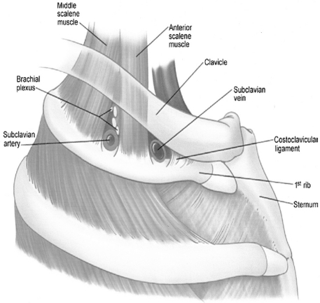
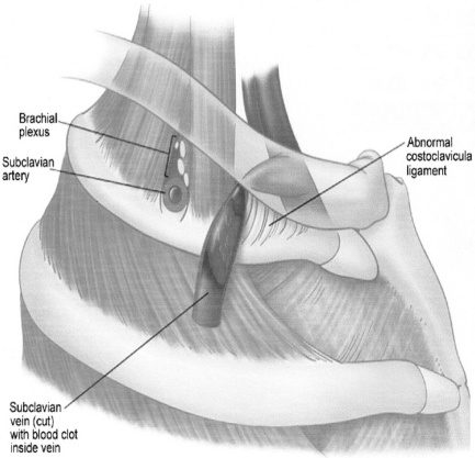
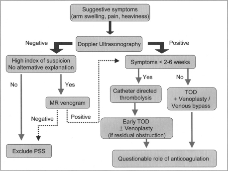
Effort thrombosis, or Paget-Schroetter Syndrome, refers to axillary-subclavian vein thrombosis associated with strenuous and repetitive activity of the upper extremities. Anatomical abnormalities at the thoracic outlet and repetitive trauma to the endothelium of the subclavian vein are key factors in its initiation and progression. The role of hereditary and acquired thrombophilias is unclear. The pathogenesis of effort thrombosis is thus distinct from other venous thromboembolic disorders. Doppler ultrasonography is the preferred initial test, while contrast venography remains the gold standard for diagnosis. Computed tomographic venography and magnetic resonance venography are comparable to conventional venography and are being increasingly used. Conservative management with anticoagulation alone is inadequate and leads to significant residual disability. An aggressive multimodal treatment strategy consisting of catheter-directed thrombolysis, with or without early thoracic outlet decompression, is essential for optimizing outcomes. Despite excellent insights into its pathogenesis and advances in treatment, a significant number of patients with effort thrombosis continue to be treated suboptimally. Hence, there is an urgent need for increasing physician awareness about risk factors, etiology and the management of this unique and relatively infrequent disorder.
Figures
References
-
- Cruveilhier LJB. Essai sur l’anatomie pathologique en général et sur les transformations et productions organiques en particulier. 1816. Doctoral thesis. Paris,
-
- Paget J. Clinical Lectures and Essays. London: Longman Green; 1875. p. 292.
-
- Von Schrötter L. Nathnagel’s Handbuch der speciellen Pathologie und Therapie. Vienna, Austria: Holder; 1884. Erkrankungen der Gefässe.
-
- Hughes ESR. Venous obstruction in the upper extremity (Paget-Schroetter’s syndrome) Br J Surg. 1948;36:155–63. - PubMed
-
- Hughes ES. Venous obstruction in the upper extremity; Paget-Schroetter’s syndrome; A review of 320 cases. Surg Gynecol Obstet. 1949;88:89–127. - PubMed