

Heterogeneous pattern of retinal nerve fiber layer in multiple sclerosis. High resolution optical coherence tomography: potential and limitations
- PMID: 21079732
- PMCID: PMC2975633
- DOI: 10.1371/journal.pone.0013877
Heterogeneous pattern of retinal nerve fiber layer in multiple sclerosis. High resolution optical coherence tomography: potential and limitations
Abstract
Background: Recently the reduction of the retinal nerve fibre layer (RNFL) was suggested to be associated with diffuse axonal damage in the whole CNS of multiple sclerosis (MS) patients. However, several points are still under discussion. (1) Is high resolution optical coherence tomography (OCT) required to detect the partly very subtle RNFL changes seen in MS patients? (2) Can a reduction of RNFL be detected in all MS patients, even in early disease courses and in all MS subtypes? (3) Does an optic neuritis (ON) or focal lesions along the visual pathways, which are both very common in MS, limit the predication of diffuse axonal degeneration in the whole CNS? The purpose of our study was to determine the baseline characteristics of clinical definite relapsing-remitting (RRMS) and secondary progressive (SPMS) MS patients with high resolution OCT technique.
Methodology: Forty-two RRMS and 17 SPMS patients with and without history of uni- or bilateral ON, and 59 age- and sex-matched healthy controls were analysed prospectively with the high resolution spectral-domain OCT device (SD-OCT) using the Spectralis 3.5mm circle scan protocol with locked reference images and eye tracking mode. Furthermore we performed tests for visual and contrast acuity and sensitivity (ETDRS, Sloan and Pelli-Robson-charts), for color vision (Lanthony D-15), the Humphrey visual field and visual evoked potential testing (VEP).
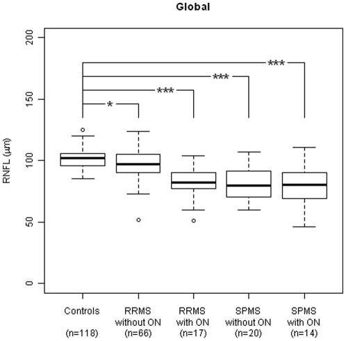
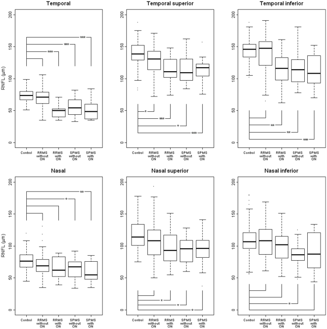
Principal findings: All 4 groups (RRMS and SPMS with or without ON) showed significantly reduced RNFL globally, or at least in one of the peripapillary sectors compared to age-/sex-matched healthy controls. In patients with previous ON additional RNFL reduction was found. However, in many RRMS patients the RNFL was found within normal range. We found no correlation between RNFL reduction and disease duration (range 9-540 months).
Conclusions: RNFL baseline characteristics of RRMS and SPMS are heterogeneous (range from normal to markedly reduced levels).
Conflict of interest statement
Figures

Similar articles
-
Relation of visual function to retinal nerve fiber layer thickness in multiple sclerosis.Ophthalmology. 2006 Feb;113(2):324-32. doi: 10.1016/j.ophtha.2005.10.040. Epub 2006 Jan 10. Ophthalmology. 2006. PMID: 16406539
-
Interpretation of RNFLT values in multiple sclerosis-associated acute optic neuritis using high-resolution SD-OCT device.Acta Ophthalmol. 2012 Sep;90(6):540-5. doi: 10.1111/j.1755-3768.2010.02013.x. Epub 2010 Nov 2. Acta Ophthalmol. 2012. PMID: 21044275
-
Evaluation of retinal nerve fiber layer thickness measured by optical coherence tomography in Moroccan patients with multiple sclerosis.J Fr Ophtalmol. 2015 Jun;38(6):497-503. doi: 10.1016/j.jfo.2014.11.008. Epub 2015 Apr 18. J Fr Ophtalmol. 2015. PMID: 25896580
-
Vision in multiple sclerosis: the story, structure-function correlations, and models for neuroprotection.J Neuroophthalmol. 2011 Dec;31(4):362-73. doi: 10.1097/WNO.0b013e318238937f. J Neuroophthalmol. 2011. PMID: 22089500 Free PMC article. Review.
-
Optical coherence tomography in optic neuritis and multiple sclerosis: a review.Eur J Neurol. 2007 Aug;14(8):841-9. doi: 10.1111/j.1468-1331.2007.01736.x. Eur J Neurol. 2007. PMID: 17662003 Review.
Cited by
-
Detection of Retinal Nerve Fiber Layer Defects in Alzheimer's Disease Using SD-OCT.Front Psychiatry. 2014 Feb 25;5:22. doi: 10.3389/fpsyt.2014.00022. eCollection 2014. Front Psychiatry. 2014. PMID: 24616709 Free PMC article.
-
Regional retinal vulnerability in multiple sclerosis: integrating OCT, MRI, and clinical data for enhanced diagnosis and automated monitoring.Rom J Morphol Embryol. 2025 Jan-Mar;66(1):119-130. doi: 10.47162/RJME.66.1.11. Rom J Morphol Embryol. 2025. PMID: 40384198 Free PMC article.
-
Longitudinal Development of Peripapillary Hyper-Reflective Ovoid Masslike Structures Suggests a Novel Pathological Pathway in Multiple Sclerosis.Ann Neurol. 2020 Aug;88(2):309-319. doi: 10.1002/ana.25782. Epub 2020 Jun 9. Ann Neurol. 2020. PMID: 32426856 Free PMC article.
-
Broadband superluminescent diode-based ultrahigh resolution optical coherence tomography for ophthalmic imaging.J Biomed Opt. 2011 Dec;16(12):126006. doi: 10.1117/1.3660314. J Biomed Opt. 2011. PMID: 22191923 Free PMC article.
-
Optical coherence tomography segmentation analysis in relapsing remitting versus progressive multiple sclerosis.PLoS One. 2017 Feb 13;12(2):e0172120. doi: 10.1371/journal.pone.0172120. eCollection 2017. PLoS One. 2017. PMID: 28192539 Free PMC article.
References
-
- Kallenbach K, Frederiksen J. Optical coherence tomography in optic neuritis and multiple sclerosis: a review. Eur J Neurol. 2007;14:841–849. - PubMed
-
- Sergott RC, Frohman E, Glanzman R, Al-Sabbagh A. The role of optical coherence tomography in multiple sclerosis: expert panel consensus. J Neurol Sci. 2007;263:3–14. - PubMed
-
- Sepulcre J, Murie-Fernandez M, Salinas-Alaman A, García-Layana A, Bejarano B, et al. Diagnostic accuracy of retinal abnormalities in predicting disease activity in MS. Neurology. 2007;68:1488–1494. - PubMed
-
- Serbecic N, Beutelspacher SC, Aboul-Enein F, Kirchner K, Reitner A. Reproducibility of High Resolution Optical Coherence Tomography Measurements of the Nerve Fibre Layer with the new Heidelberg Spectralis OCT. Br J Ophthalmol. 2010 accepted (MEDLINE INDEX MISSING, EXPECTED IN NOVEMBER 2010) - PubMed
-
- Henderson AP, Trip SA, Schlottmann PG, Altmann DR, Garway-Heath DF, et al. A preliminary longitudinal study of the retinal nerve fiber layer in progressive multiple sclerosis. J Neurol. 2010;257:1083–1091. - PubMed
Publication types
MeSH terms
LinkOut - more resources
Full Text Sources
Medical
Miscellaneous
