. 2010 Nov 5;5(11):e13854.
doi: 10.1371/journal.pone.0013854.

Proteomic analysis of the dysferlin protein complex unveils its importance for sarcolemmal maintenance and integrity

Affiliations

Proteomic analysis of the dysferlin protein complex unveils its importance for sarcolemmal maintenance and integrity

Antoine de Morrée et al. PLoS One. .

Abstract

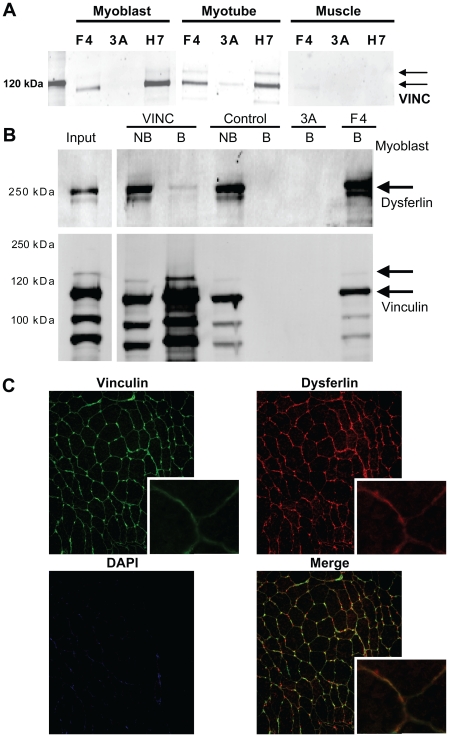
Dysferlin is critical for repair of muscle membranes after damage. Mutations in dysferlin lead to a progressive muscular dystrophy. Recent studies suggest additional roles for dysferlin. We set out to study dysferlin's protein-protein interactions to obtain comprehensive knowledge of dysferlin functionalities in a myogenic context. We developed a robust and reproducible method to isolate dysferlin protein complexes from cells and tissue. We analyzed the composition of these complexes in cultured myoblasts, myotubes and skeletal muscle tissue by mass spectrometry and subsequently inferred potential protein functions through bioinformatics analyses. Our data confirm previously reported interactions and support a function for dysferlin as a vesicle trafficking protein. In addition novel potential functionalities were uncovered, including phagocytosis and focal adhesion. Our data reveal that the dysferlin protein complex has a dynamic composition as a function of myogenic differentiation. We provide additional experimental evidence and show dysferlin localization to, and interaction with the focal adhesion protein vinculin at the sarcolemma. Finally, our studies reveal evidence for cross-talk between dysferlin and its protein family member myoferlin. Together our analyses show that dysferlin is not only a membrane repair protein but also important for muscle membrane maintenance and integrity.


Conflict of interest statement

Competing Interests: The authors have declared that no competing interests exist.

Figures

Figure 1
Figure 1. Dysferlin is upregulated during myogenic differentiation in IM2 cells.
IM2 cells were differentiated and harvested at day 0, 1, 3 and 5. Protein lysates were probed on western blot for Dysferlin (upper panel) and Calpain 3 (lower panel).
Figure 2
Figure 2. Reproducible dysferlin immunoprecipitation under different conditions.
IM2 myoblasts and myotubes were lysed in three different buffers and subjected to a HCAb Dysferlin immuno precipitation protocol. F4 and H7 are specific for Dysferlin while 3A is a non-specfic control HCAb. A) Coomassie stained gel of immunoprecipitation fractions from myoblasts. B) western blot for Dysferlin and Calpain 3 corresponding to the gel in A. C) A similar western blot on myotubes IP fractions (corresponding gel not shown).
Figure 3
Figure 3. Immunoprecipitation of dysferlin from different myogenic sources is highly reproducible.
A) Coomassie blue stained gel of IP fractions from IM2 myoblasts, myotubes and skeletal muscle tissue. IP samples of two dysferlin specific HCAb (F4 and H7) yield highly similar staining patterns, contrary to a non-specific HCAb (3A). B) a concomitant western blot for dysferlin confirms the IP. C) All protein bands stained in A), lanes H7, were excised, in-gel trypsin digested and analyzed by mass spectrometry. Many described interaction partners of dysferlin were identified by western blot (Figure S2) and thus confirmed.
Figure 4
Figure 4. Venn diagram showing the overlap of dysferlin interaction partners in myoblast, myotubes and skeletal muscle tissue.
All Coomassie stained bands from the H7 IP shown in Figure 3A were excised, in-gel digested and analyzed by mass spectrometry. 115 proteins are consistently identified in all three protein sources.
Figure 5
Figure 5. Myoferlin and dysferlin may be present in the same protein complex.
A) Dysferlin IP fractions were stained in western blot with a myoferlin specific antibody, to verify that full-length dysferlin is co-immunoprecipitated with dysferlin, and not with the control HCAb 3A. B) U2OS cells, which express endogenous myoferlin but not dysferlin, were transfected with dysferlin cDNA or empty vector (mock), and both untransfected (wild-type) and transfected cells were lysed and subjected to an IP experiment with F4 and H7. IP and non-bound fractions were analyzed on western blot for dysferlin (lower panel) and myoferlin (upper panel). F4 and H7 can only immunoprecipitate dysferlin from dysferlin transfected cells (lower panel), consistent with the absence of endogenous dysferlin expression. Myoferlin is not immunoprecipitated from wild-type cells, though it can be detected in the non-bound fractions. However, myoferlin is immunoprecipitated by F4 from dysferlin expressing U2OS cells (band marked by asterisk).
Figure 6
Figure 6. A potential interaction between dysferlin and vinculin.
A) The dysferlin IP samples were analyzed on western blot for vinculin, to verify that full-length vinculin specifically co-immunoprecipitates with dysferlin, but not with the 3A control HCAb. B) cell lysate of IM2 myotubes were subjected to immunoprecipitate with antibodies against vinculin and an IgG control (against VSV), and IP and non-bound fractions were analyzed for dysferlin on western blot. HCAb F4 and 3A were taken along as positive and negative controls, respectively. Dysferlin is strongly detected in the F4 fraction (upper panel). Weak signal is also seen in the vinculin bound fractions, indicating a weak interaction with dysferlin. A corresponding western blot for vinculin shows the presence of full-length vinculin in the F4 and vinculin IP fractions (lower panel). C) Human skeletal muscle cryosections were stained for vinculin (green) and dysferlin (F4, red). In cross-sections (right) both dysferlin and vinculin are detected at the sarcolemma.

Similar articles

Cited by

References

    1. Anderson LV, Davison K, Moss JA, Young C, Cullen MJ, et al. Dysferlin is a plasma membrane protein and is expressed early in human development. Hum Mol Genet. 1999;8:855–861. ddc087 [pii] - PubMed
    1. Bansal D, Miyake K, Vogel SS, Groh S, Chen CC, et al. Defective membrane repair in dysferlin-deficient muscular dystrophy. Nature. 2003;423:168–172. 10.1038/nature01573 [doi];nature01573 [pii] - PubMed
    1. Bashir R, Britton S, Strachan T, Keers S, Vafiadaki E, et al. A gene related to Caenorhabditis elegans spermatogenesis factor fer-1 is mutated in limb-girdle muscular dystrophy type 2B. Nat Genet. 1998;20:37–42. 10.1038/1689 [doi] - PubMed
    1. Illa I, Serrano-Munuera C, Gallardo E, Lasa A, Rojas-Garcia R, et al. Distal anterior compartment myopathy: a dysferlin mutation causing a new muscular dystrophy phenotype. Ann Neurol. 2001;49:130–134. - PubMed
    1. Liu J, Aoki M, Illa I, Wu C, Fardeau M, et al. Dysferlin, a novel skeletal muscle gene, is mutated in Miyoshi myopathy and limb girdle muscular dystrophy. Nat Genet. 1998;20:31–36. 10.1038/1682 [doi] - PubMed

Publication types

MeSH terms