. 2010 Nov 5;5(11):e13858.
doi: 10.1371/journal.pone.0013858.

Changes in hepatic gene expression upon oral administration of taurine-conjugated ursodeoxycholic acid in ob/ob mice

Affiliations

Changes in hepatic gene expression upon oral administration of taurine-conjugated ursodeoxycholic acid in ob/ob mice

Jae-Seong Yang et al. PLoS One. .

Abstract

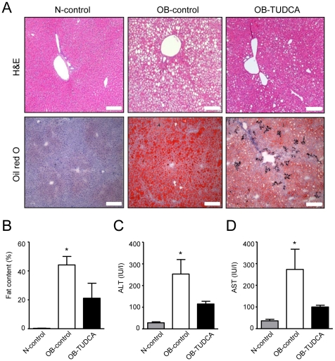
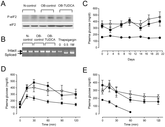
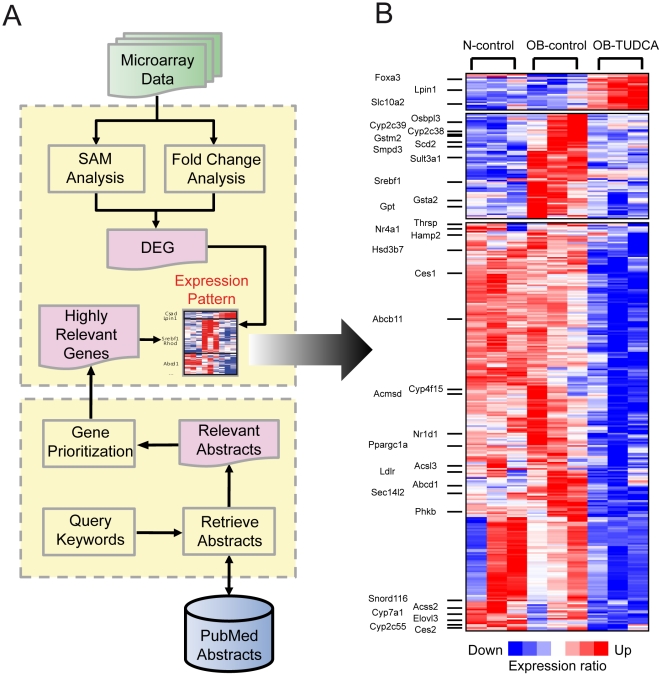
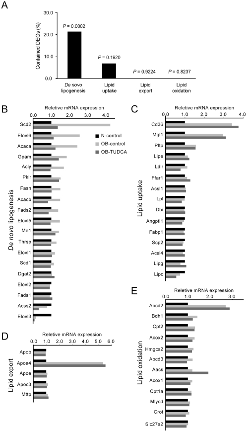
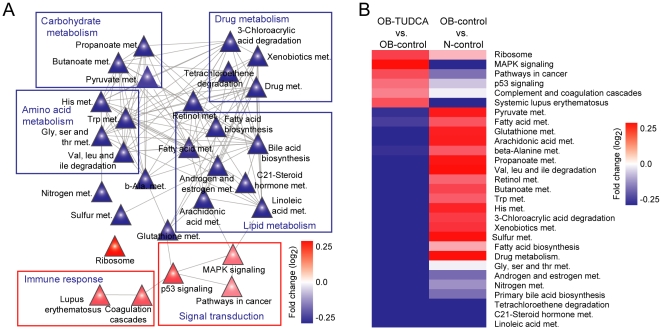
Nonalcoholic fatty liver disease (NAFLD) is highly prevalent and associated with considerable morbidities. Unfortunately, there is no currently available drug established to treat NAFLD. It was recently reported that intraperitoneal administration of taurine-conjugated ursodeoxycholic acid (TUDCA) improved hepatic steatosis in ob/ob mice. We hereby examined the effect of oral TUDCA treatment on hepatic steatosis and associated changes in hepatic gene expression in ob/ob mice. We administered TUDCA to ob/ob mice at a dose of 500 mg/kg twice a day by gastric gavage for 3 weeks. Body weight, glucose homeostasis, endoplasmic reticulum (ER) stress, and hepatic gene expression were examined in comparison with control ob/ob mice and normal littermate C57BL/6J mice. Compared to the control ob/ob mice, TUDCA treated ob/ob mice revealed markedly reduced liver fat stained by oil red O (44.2±5.8% vs. 21.1±10.4%, P<0.05), whereas there was no difference in body weight, oral glucose tolerance, insulin sensitivity, and ER stress. Microarray analysis of hepatic gene expression demonstrated that oral TUDCA treatment mainly decreased the expression of genes involved in de novo lipogenesis among the components of lipid homeostasis. At pathway levels, oral TUDCA altered the genes regulating amino acid, carbohydrate, and drug metabolism in addition to lipid metabolism. In summary, oral TUDCA treatment decreased hepatic steatosis in ob/ob mice by cooperative regulation of multiple metabolic pathways, particularly by reducing the expression of genes known to regulate de novo lipogenesis.


Conflict of interest statement

Competing Interests: Y.M.C. received a research grant from Daewoong Co. LTD., Seoul, South Korea. Daewoong Co. LTD. is producing and marketing ursodeoxycholic acid. The authors confirm that this does not alter their adherence to all the PLoS ONE policies on sharing data and materials.

Figures

Figure 1
Figure 1. Effects of oral TUDCA treatment on hepatic steatosis in ob/ob mice.
(A) H&E and oil red O staining of the liver shows a marked improvement of steatosis. Scale bars indicate 200 µm. (B) Quantified fat content in oil red O stained liver sections was lower in the OB-TUDCA group as compared to the OB-control group. (C, D) Serum ALT and AST levels tended to be lower in the OB-TUDCA group as compared to the OB-control group. In panels B, C and D, * denotes P<0.05 compared to the N-control. P-values were calculated using the Kruskal-Wallis test and Dunn's multiple comparison test.
Figure 2
Figure 2. Effects of oral TUDCA treatment on lipid peroxidation.
(A) The 4-HNE staining of the liver shows that lipid peroxidation is increased in ob/ob mice, which is reduced by TUDCA treatment. Scale bars in the top and bottom images indicate 200 µm and 800 µm, respectively. (B) Quantification of the intensity of 4-HNE staining. * denotes P<0.05 compared to the N-control, and # denotes P<0.05 compared to OB-control.
Figure 3
Figure 3. Effects of oral TUDCA treatment on ER stress and glucose homeostasis in ob/ob mice.
(A) Phosphorylated eIF2α expression levels are similar among groups. (B) Alternative splicing of Xbp1 was not detected in any of the three groups. The mRNAs isolated from thapsigargin-treated 3T3 L1 cells were used as controls. (C–E) There was no difference in fed-state blood glucose levels (C), glucose tolerance assessed by IPGTT (D), and insulin sensitivity assessed by ITT (E) between the OB-control and OB-TUDCA groups. Symbols and error bars represent means and SEM, respectively. (•), N-control; (○), OB-control; and (▪), OB-TUDCA mice.
Figure 4
Figure 4. Changes in hepatic gene expression in OB-control and OB-TUDCA groups.
(A) Flowchart of DEG selection and relevant gene search. DEGs were selected using SAM and fold-change analysis. The relevant genes were selected using a PubMed database search. (B) The expression matrix shows the relative expression levels of DEGs for each condition. Each column of the matrix represents three experimental conditions: N-control, OB-control, and OB-TUDCA groups. Each row of the matrix represents relative expression data that were gene-wise normalized. Hierarchical clustering was conducted using the following parameters: Pearson correlation and single linkage method.
Figure 5
Figure 5. Alteration of gene expression in lipid flux regulating groups.
(A) The percentages of included DEGs in each group and its statistical significance. (B–E) Expression changes of genes involved in de novo lipogenesis, lipid uptake, export, and oxidation.
Figure 6
Figure 6. Relationships between altered pathways in the OB-TUDCA group.
(A) Abstract view of the pathway-pathway network; it shows the relationship among the pathways by connecting common genes. The colors of triangles represent the average changes in expression of genes involved in the pathway. (B) Average log2 fold-change expression pattern of DEPs in N-control vs. OB-control groups and OB-TUDCA vs. OB-control groups. Hierarchical clustering was conducted using the following parameters: Pearson correlation and single linkage method.

References

    1. Angulo P. Nonalcoholic fatty liver disease. N Engl J Med. 2002;346:1221–1231. - PubMed
    1. Greenfield V, Cheung O, Sanyal AJ. Recent advances in nonalcholic fatty liver disease. Curr Opin Gastroenterol. 2008;24:320–327. - PubMed
    1. Stefan N, Kantartzis K, Haring HU. Causes and metabolic consequences of Fatty liver. Endocr Rev. 2008;29:939–960. - PubMed
    1. Browning JD, Szczepaniak LS, Dobbins R, Nuremberg P, Horton JD, et al. Prevalence of hepatic steatosis in an urban population in the United States: impact of ethnicity. Hepatology. 2004;40:1387–1395. - PubMed
    1. Targher G, Bertolini L, Rodella S, Tessari R, Zenari L, et al. Nonalcoholic fatty liver disease is independently associated with an increased incidence of cardiovascular events in type 2 diabetic patients. Diabetes Care. 2007;30:2119–2121. - PubMed

Publication types

MeSH terms