. 2010 Nov 4;5(11):e13839.
doi: 10.1371/journal.pone.0013839.

Antioxidants protect keratinocytes against M. ulcerans mycolactone cytotoxicity

Affiliations

Antioxidants protect keratinocytes against M. ulcerans mycolactone cytotoxicity

Alvar Grönberg et al. PLoS One. .

Abstract

Background: Mycobacterium ulcerans is the causative agent of necrotizing skin ulcerations in distinctive geographical areas. M. ulcerans produces a macrolide toxin, mycolactone, which has been identified as an important virulence factor in ulcer formation. Mycolactone is cytotoxic to fibroblasts and adipocytes in vitro and has modulating activity on immune cell functions. The effect of mycolactone on keratinocytes has not been reported previously and the mechanism of mycolactone toxicity is presently unknown. Many other macrolide substances have cytotoxic and immunosuppressive activities and mediate some of their effects via production of reactive oxygen species (ROS). We have studied the effect of mycolactone in vitro on human keratinocytes--key cells in wound healing--and tested the hypothesis that the cytotoxic effect of mycolactone is mediated by ROS.

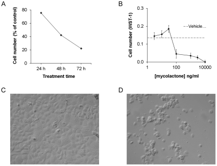
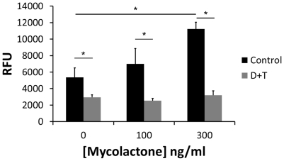
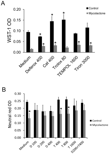
Methodology/principal findings: The effect of mycolactone on primary skin keratinocyte growth and cell numbers was investigated in serum free growth medium in the presence of different antioxidants. A concentration and time dependent reduction in keratinocyte cell numbers was observed after exposure to mycolactone. Several different antioxidants inhibited this effect partly. The ROS inhibiting substance deferoxamine, which acts via chelation of Fe(2+), completely prevented mycolactone mediated cytotoxicity.

Conclusions/significance: This study demonstrates that mycolactone mediated cytotoxicity can be inhibited by deferoxamine, suggesting a role of iron and ROS in mycolactone induced cytotoxicity of keratinocytes. The data provide a basis for the understanding of Buruli ulcer pathology and the development of improved therapies for this disease.


Conflict of interest statement

Competing Interests: A.G. is and L.Z. was employed by Lipopeptide AB (Stockholm, Sweden). M.S. and J.H. are founders and shareholders of Lipopeptide AB. This does not alter the authors' adherence to all the PLoS ONE policies on sharing data and materials. Lipopeptide AB is developing a wound healing product intended for hard-to-heal venous leg ulcers (www.lipopeptide.se). The company has no products on the market. Lipopeptide gave limited support to this work but has denied rights regarding the work presented in this manuscript to A.G. No patent has been filed on work involving research on Buruli ulcers by the company or its employees.

Figures

Figure 1
Figure 1. Mycolactone is cytotoxic for keratinocytes.
Mycolactone was added to sub-confluent cultures of keratinocytes and (A) cell numbers were measured as optical density of WST-1after 24, 48 and 72 h of treatment with 1000 ng/ml of mycolactone, or (B) after 72 h at different concentrations of mycolactone. An asterisk indicates a significant (p<0.05) reduction in WST-1 labeling as compared to the corresponding medium control. Cell cultures of untreated controls (C) and cultures treated with 300 ng/m mycolactone (D) were photographed after 48 h. Data are from one representative experiment showing means and SD of triplicates.
Figure 2
Figure 2. Mycolactone increases ROS production.
Intracellular ROS production was detected by intracellular CM-H2DCFDA fluorescence in keratinocytes after 45 min incubation in control medium or with 100 and 300 ng/ml mycolactone. Parallel cultures were pretreated for 30 min with a combination of deferoxamine (100 µM) and TEMPOL (200 µM) (D+T) before CM-H2DCFDA and addition of mycolactone. Asterisks indicate significant (p<0.05) differences in fluorescence between treatments and their corresponding control in one representative experiment with four to six replicates (means and SD).
Figure 3
Figure 3. Antioxidant protection against mycolactone cytotoxicity.
(A) Antioxidants were added 30 min before mycolactone (300 ng/ml) and the incubation was continued for 48 h when cell numbers were determined by measuring WST-1color at 450 nm. Data represent means of triplicate determinations and standard deviations from one representative experiment out of two performed (*, p<0.05, one way ANOVA and students t-test). (B) Deferoxamine (D) and TEMPOL were added at different concentrations alone or in combination 30 min before mycolactone (300 ng/ml) and the incubation was continued for 48 h when cell numbers were determined by measuring neutral red uptake Concentrations are in µM for all substances except catalase which is in U/ml. Data shown are means and SEM of four experiments (*, p<0.05, ANOVA and students t-test). TEMPOL 1600 µM was not included in the statistical analysis (N = 2).

References

    1. George KM, Chatterjee D, Gunawardana G, Welty D, Hayman J, et al. Mycolactone: a polyketide toxin from Mycobacterium ulcerans required for virulence. Science. 1999;283:854–857. - PubMed
    1. Hong H, Demangel C, Pidot SJ, Leadlay PF, Stinear T. Mycolactones: immunosuppressive and cytotoxic polyketides produced by aquatic mycobacteria. Nat Prod Rep. 2008;25:447–454. - PMC - PubMed
    1. Mve-Obiang A, Lee RE, Portaels F, Small PL. Heterogeneity of mycolactones produced by clinical isolates of Mycobacterium ulcerans: implications for virulence. Infect Immun. 2003;71:774–783. - PMC - PubMed
    1. Adusumilli S, Mve-Obiang A, Sparer T, Meyers W, Hayman J, et al. Mycobacterium ulcerans toxic macrolide, mycolactone modulates the host immune response and cellular location of M. ulcerans in vitro and in vivo. Cell Microbiol. 2005;7:1295–1304. - PubMed
    1. George KM, Pascopella L, Welty DM, Small PL. A Mycobacterium ulcerans toxin, mycolactone, causes apoptosis in guinea pig ulcers and tissue culture cells. Infect Immun. 2000;68:877–883. - PMC - PubMed

MeSH terms