

TXNL6 is a novel oxidative stress-induced reducing system for methionine sulfoxide reductase a repair of α-crystallin and cytochrome C in the eye lens
- PMID: 21079812
- PMCID: PMC2973970
- DOI: 10.1371/journal.pone.0015421
TXNL6 is a novel oxidative stress-induced reducing system for methionine sulfoxide reductase a repair of α-crystallin and cytochrome C in the eye lens
Abstract
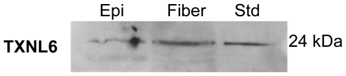
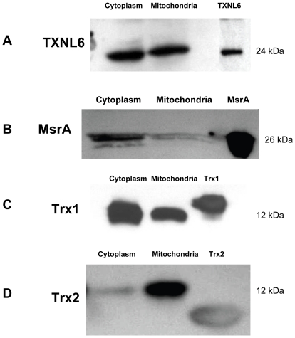
A key feature of many age-related diseases is the oxidative stress-induced accumulation of protein methionine sulfoxide (PMSO) which causes lost protein function and cell death. Proteins whose functions are lost upon PMSO formation can be repaired by the enzyme methionine sulfoxide reductase A (MsrA) which is a key regulator of longevity. One disease intimately associated with PMSO formation and loss of MsrA activity is age-related human cataract. PMSO levels increase in the eye lens upon aging and in age-related human cataract as much as 70% of total lens protein is converted to PMSO. MsrA is required for lens cell maintenance, defense against oxidative stress damage, mitochondrial function and prevention of lens cataract formation. Essential for MsrA action in the lens and other tissues is the availability of a reducing system sufficient to catalytically regenerate active MsrA. To date, the lens reducing system(s) required for MsrA activity has not been defined. Here, we provide evidence that a novel thioredoxin-like protein called thioredoxin-like 6 (TXNL6) can serve as a reducing system for MsrA repair of the essential lens chaperone α-crystallin/sHSP and mitochondrial cytochrome c. We also show that TXNL6 is induced at high levels in human lens epithelial cells exposed to H(2)O(2)-induced oxidative stress. Collectively, these data suggest a critical role for TXNL6 in MsrA repair of essential lens proteins under oxidative stress conditions and that TXNL6 is important for MsrA defense protection against cataract. They also suggest that MsrA uses multiple reducing systems for its repair activity that may augment its function under different cellular conditions.
Conflict of interest statement
Figures


Similar articles
-
Methionine sulfoxide reductase A (MsrA) restores alpha-crystallin chaperone activity lost upon methionine oxidation.Biochim Biophys Acta. 2009 Dec;1790(12):1665-72. doi: 10.1016/j.bbagen.2009.08.011. Epub 2009 Sep 3. Biochim Biophys Acta. 2009. PMID: 19733220 Free PMC article.
-
Deletion of mouse MsrA results in HBO-induced cataract: MsrA repairs mitochondrial cytochrome c.Mol Vis. 2009 May 15;15:985-99. Mol Vis. 2009. PMID: 19461988 Free PMC article.
-
Methionine sulfoxide reductase A is important for lens cell viability and resistance to oxidative stress.Proc Natl Acad Sci U S A. 2004 Jun 29;101(26):9654-9. doi: 10.1073/pnas.0403532101. Epub 2004 Jun 15. Proc Natl Acad Sci U S A. 2004. PMID: 15199188 Free PMC article.
-
Mitochondrial function and redox control in the aging eye: role of MsrA and other repair systems in cataract and macular degenerations.Exp Eye Res. 2009 Feb;88(2):195-203. doi: 10.1016/j.exer.2008.05.018. Epub 2008 Jun 7. Exp Eye Res. 2009. PMID: 18588875 Free PMC article. Review.
-
Structural and functional properties, chaperone activity and posttranslational modifications of alpha-crystallin and its related subunits in the crystalline lens: N-acetylcarnosine, carnosine and carcinine act as alpha- crystallin/small heat shock protein enhancers in prevention and dissolution of cataract in ocular drug delivery formulations of novel therapeutic agents.Recent Pat Drug Deliv Formul. 2012 Aug;6(2):107-48. doi: 10.2174/187221112800672976. Recent Pat Drug Deliv Formul. 2012. PMID: 22436026 Review.
Cited by
-
Parkin elimination of mitochondria is important for maintenance of lens epithelial cell ROS levels and survival upon oxidative stress exposure.Biochim Biophys Acta Mol Basis Dis. 2017 Jan;1863(1):21-32. doi: 10.1016/j.bbadis.2016.09.020. Epub 2016 Oct 1. Biochim Biophys Acta Mol Basis Dis. 2017. PMID: 27702626 Free PMC article.
-
Metabolic and Redox Signaling of the Nucleoredoxin-Like-1 Gene for the Treatment of Genetic Retinal Diseases.Int J Mol Sci. 2020 Feb 27;21(5):1625. doi: 10.3390/ijms21051625. Int J Mol Sci. 2020. PMID: 32120883 Free PMC article. Review.
-
Viral-mediated RdCVF and RdCVFL expression protects cone and rod photoreceptors in retinal degeneration.J Clin Invest. 2015 Jan;125(1):105-16. doi: 10.1172/JCI65654. Epub 2014 Nov 21. J Clin Invest. 2015. PMID: 25415434 Free PMC article.
-
Optimal Control with RdCVFL for Degenerating Photoreceptors.Bull Math Biol. 2024 Feb 12;86(3):29. doi: 10.1007/s11538-024-01256-6. Bull Math Biol. 2024. PMID: 38345678 Free PMC article.
-
Metabolic and redox signaling in the retina.Cell Mol Life Sci. 2017 Oct;74(20):3649-3665. doi: 10.1007/s00018-016-2318-7. Epub 2016 Aug 20. Cell Mol Life Sci. 2017. PMID: 27543457 Free PMC article. Review.
References
-
- Gabbita SP, Aksenov MY, Lovell MA, Markesbery WR. Decrease in peptide methionine sulfoxide reductase in Alzheimer's disease brain. Journal of Neurochemistry. 1999;73:1660–1666. - PubMed
-
- Schoneich C. Methionine oxidation by reactive oxygen species: reaction mechanisms and relevance to Alzheimer's disease. Biochemica et Biophyisca Acta. 2005;1703:111–119. doi: 10.1016/j.bbapap.2004.09.009. - DOI - PubMed
-
- Glaser CB, Yamin G, Uversky VN, Fink AL. Methionine oxidation, alpha-synuclein and Parkinson's disease. Biochimica et Biophysica Acta. 2005;1703:157–169. doi: 10.1016/j.bbapap.2004.10.008. - DOI - PubMed
-
- Liu F, Hindupur J, Nguyen JL, Ruf KJ, Zhu J, et al. Methionine sulfoxide reductase A protects dopaminergic cells from Parkinson's disease-related insults. Free Radical Biology and Medicine. 2008;45:242–255. doi: 10.1016/j.freeradbiomed.2008.03.022. - DOI - PMC - PubMed
-
- Wassef R, Haenold R, Hansel A, Brot N, Heinemann SH, et al. Methionine sulfoxide reductase A and a dietary supplement S-methyl-L-cysteine prevent Parkinson's-like symptoms. Neuroscience. 2007;27:12808–12816. doi: 10.1523/JNEUROSCI.0322-07.2007. - DOI - PMC - PubMed
Publication types
MeSH terms
Substances
Grants and funding
LinkOut - more resources
Full Text Sources
Other Literature Sources
Molecular Biology Databases
