Review
. 2011 Apr;89(4):321-9.
doi: 10.1007/s00109-010-0697-z. Epub 2010 Nov 16.

Hematopoietic colony-stimulating factors: new players in tumor-nerve interactions

Affiliations
Review

Hematopoietic colony-stimulating factors: new players in tumor-nerve interactions

Sebastian Stösser et al. J Mol Med (Berl). 2011 Apr.

Abstract

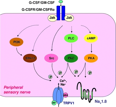
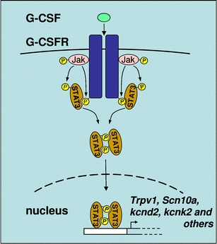
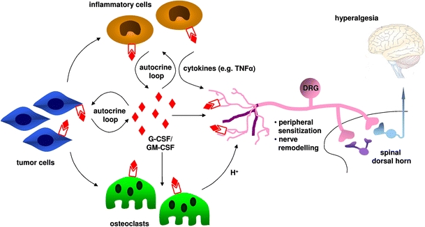
A variety of cancers are accompanied by debilitating pain, which constitutes the primary reason for poor quality of life in cancer patients. There is an urgent demand for the development of specific mechanism-based therapies against cancer pain. Recently, important advances have been made in mechanisms contributing to cancer pain. A notable finding was that the tumor-derived hematopoietic growth factors, granulocyte- and granulocyte-macrophage-colony-stimulating factors (G-CSF/GM-CSF), subserve important functions in the generation of pain hypersensitivity in tumor-affected regions. In this context, their receptors were unexpectedly found on pain-sensing nerves and were observed to be functionally linked to nociceptive sensitization and tumor-induced pain. Here, we review evidence supporting a role for G-/GM-CSF in sensitization of pain-sensing nerves, the underlying signaling pathways and the cross-talk with other pronociceptive cytokines, peptides and modulators derived from immune cells, osteoclasts and tumor cells. These findings hold implications in the therapy of pain in disease states, such as cancer and rheumatoid arthritis.


Figures

Fig. 1
Fig. 1
Schematic representation of potential signaling mechanisms linking transmembrane receptors for G-CSF and GM-CSF to sensitization of nociceptive transducers in peripheral sensory neurons. Binding of G-CSF or GM-CSF to their cognate transmembrane receptors can activate a myriad of signaling cascades resulting ultimately in sensitization and increased membrane levels of TRPV1 and Nav1.8, which representatively depicts the process of nociceptor sensitization here. TRPV1 transduces heat, protons and lipids, such as anandamide (AEA) and Nav1.8 is a key determinant of nociceptor excitability. The MAP kinases ERK1/2, Src kinases, protein kinase C (PKC) and protein kinase A (PKA) modify either the function of TRPV1 or its membrane localisation via phosphorylation. Signaling cascades stimulated differentially by G-CSF and GM-CSF are not depicted separately here. Dotted arrow lines indicate that intermediate steps of the signaling cascade were omitted in the representation
Fig. 2
Fig. 2
Further implications of G-/GM-CSF-induced JAK-STAT signaling in peripheral sensory neurons. G-/GM-CSF induces nuclear translocation of STAT3 and promotes the transcription of genes encoding TRPV1 (Trpv1), Nav1.8 (Scn10a), Kv4.2 (kcnd2), TREK-1 (kcnk2) and possibly other pain-related genes via the JAK-STAT pathway
Fig. 3
Fig. 3
Representation of potential local effects of G-CSF and GM-CSF in the micromilieu of bone metastases in the context of cancer pain. G-CSF and GM-CSF are released by tumor cells and inflammatory cells and can act on peripheral endings of dorsal root ganglion neurons to produce sensitization, which is perceived as exaggerated pain (hyperalgesia). Furthermore, G-CSF and GM-CSF can evoke nerve remodelling (hypertrophy, sprouting of sensory nerves). Additionally, G-CSF and GM-CSF can recruit and stimulate inflammatory cells and induce bone resorption by osteoclasts. Inflammatory cells themselves sensitize peripheral nerve fibers by release of proinflammatory cytokines (e.g. TNF-α, interleukins and endothelins), which may also contribute to nerve remodelling. Furthermore, osteoclasts can contribute to peripheral sensitization, in particular by release of protons. The proliferation of some types of tumor cells can be stimulated by local G-CSF and GM-CSF. In turn, tumor cells can contribute also indirectly to the sensitization of primary afferent nerve fibers by stimulating osteolysis and the local inflammatory response. Thus, hematopoietic growth factors may serve as central players in cancer pain via diverse, parallel mechanisms

References

    1. Portenoy RK, Lesage P. Management of cancer pain. Lancet. 1999;353:1695–1700. doi: 10.1016/S0140-6736(99)01310-0. - DOI - PubMed
    1. Mantyh PW. Cancer pain and its impact on diagnosis, survival and quality of life. Nat Rev Neurosci. 2006;7:797–809. doi: 10.1038/nrn1914. - DOI - PubMed
    1. Mantyh PW, Hunt SP. Mechanisms that generate and maintain bone cancer pain. Novartis Found Symp. 2004;260:221–238. doi: 10.1002/0470867639.ch15. - DOI - PubMed
    1. Wacnik PW, Eikmeier LJ, Ruggles TR, Ramnaraine ML, Walcheck BK, Beitz AJ, Wilcox GL. Functional interactions between tumor and peripheral nerve: morphology, algogen identification, and behavioral characterization of a new murine model of cancer pain. J Neurosci. 2001;21:9355–9366. - PMC - PubMed
    1. Hamilton JA. Colony-stimulating factors in inflammation and autoimmunity. Nat Rev Immunol. 2008;8:533–544. doi: 10.1038/nri2356. - DOI - PubMed

Publication types

MeSH terms

Substances