

Clinical translation of ex vivo sentinel lymph node mapping for colorectal cancer using invisible near-infrared fluorescence light
- PMID: 21080086
- PMCID: PMC3052497
- DOI: 10.1245/s10434-010-1426-0
Clinical translation of ex vivo sentinel lymph node mapping for colorectal cancer using invisible near-infrared fluorescence light
Abstract
Background: Sentinel lymph node (SLN) mapping in colorectal cancer may have prognostic and therapeutic significance; however, currently available techniques are not optimal. We hypothesized that the combination of invisible near-infrared (NIR) fluorescent light and ex vivo injection could solve remaining problems of SLN mapping in colorectal cancer.
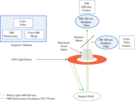
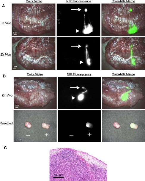
Methods: The FLARE imaging system was used for real-time identification of SLNs after injection of the NIR lymphatic tracer HSA800 in the colon and rectum of (n = 4) pigs. A total of 32 SLN mappings were performed in vivo and ex vivo after oncologic resection using an identical injection technique. Guided by these results, SLN mappings were performed in ex vivo tissue specimens of 24 consecutive colorectal cancer patients undergoing resection.
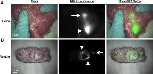
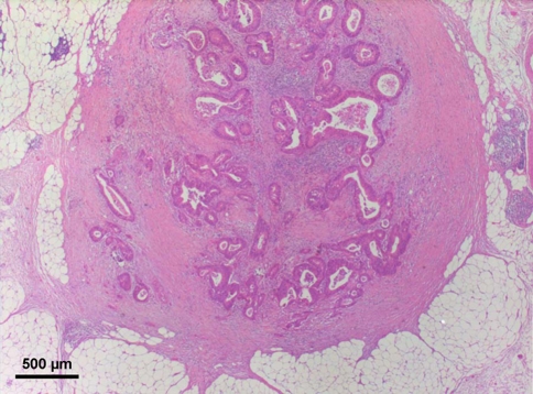
Results: Lymph flow could be followed in real-time from the injection site to the SLN using NIR fluorescence. In pigs, the SLN was identified in 32 of 32 (100%) of SLN mappings under both in vivo and ex vivo conditions. Clinically, SLNs were identified in all patients (n = 24) using the ex vivo strategy within 5 min after injection of fluorescent tracer. Also, 9 patients showed lymph node involvement (N1 disease). In 1 patient, a 3-mm mesenteric metastasis was found adjacent to a tumor-negative SLN.
Conclusions: The current pilot study shows proof of principle that ex vivo NIR fluorescence-guided SLN mapping can provide high-sensitivity, rapid, and accurate identification of SLNs in colon and rectum. This creates an experimental platform to test optimized, non-FDA-approved NIR fluorescent lymphatic tracers in a clinical setting.
Conflict of interest statement
All FLARE
Figures
Similar articles
-
Perioperative PET/CT lymphoscintigraphy and fluorescent real-time imaging for sentinel lymph node mapping in early staged colon cancer.Eur J Nucl Med Mol Imaging. 2019 Jul;46(7):1495-1505. doi: 10.1007/s00259-019-04284-w. Epub 2019 Feb 23. Eur J Nucl Med Mol Imaging. 2019. PMID: 30798428 Free PMC article.
-
Identification of metastatic nodal disease in a phase 1 dose-escalation trial of intraoperative sentinel lymph node mapping in non-small cell lung cancer using near-infrared imaging.J Thorac Cardiovasc Surg. 2013 Sep;146(3):562-70; discussion 569-70. doi: 10.1016/j.jtcvs.2013.04.010. Epub 2013 Jun 19. J Thorac Cardiovasc Surg. 2013. PMID: 23790404 Free PMC article. Clinical Trial.
-
Image-guided oncologic surgery using invisible light: completed pre-clinical development for sentinel lymph node mapping.Ann Surg Oncol. 2006 Dec;13(12):1671-81. doi: 10.1245/s10434-006-9194-6. Epub 2006 Sep 29. Ann Surg Oncol. 2006. PMID: 17009138 Free PMC article.
-
Diagnostic value of near-infrared or fluorescent indocyanine green guided sentinel lymph node mapping in gastric cancer: A systematic review and meta-analysis.J Surg Oncol. 2018 Dec;118(8):1243-1256. doi: 10.1002/jso.25285. Epub 2018 Oct 31. J Surg Oncol. 2018. PMID: 30380146
-
Sentinel node mapping with indocyanine green and endoscopic near-infrared fluorescence imaging in endometrial cancer. A pilot study and review of the literature.Gynecol Oncol. 2015 Jun;137(3):443-7. doi: 10.1016/j.ygyno.2015.03.004. Epub 2015 Mar 11. Gynecol Oncol. 2015. PMID: 25771495 Review.
Cited by
-
Dendritic polyglycerolsulfate near infrared fluorescent (NIRF) dye conjugate for non-invasively monitoring of inflammation in an allergic asthma mouse model.PLoS One. 2013;8(2):e57150. doi: 10.1371/journal.pone.0057150. Epub 2013 Feb 21. PLoS One. 2013. PMID: 23437332 Free PMC article.
-
Charge and hydrophobicity effects of NIR fluorophores on bone-specific imaging.Theranostics. 2015 Mar 1;5(6):609-17. doi: 10.7150/thno.11222. eCollection 2015. Theranostics. 2015. PMID: 25825600 Free PMC article.
-
Fluorescence Guidance in Surgical Oncology: Challenges, Opportunities, and Translation.Mol Imaging Biol. 2019 Apr;21(2):200-218. doi: 10.1007/s11307-018-1239-2. Mol Imaging Biol. 2019. PMID: 29942988 Free PMC article. Review.
-
Effects of an Unlabeled Loading Dose on Tumor-Specific Uptake of a Fluorescently Labeled Antibody for Optical Surgical Navigation.Mol Imaging Biol. 2017 Aug;19(4):610-616. doi: 10.1007/s11307-016-1022-1. Mol Imaging Biol. 2017. PMID: 27830425 Free PMC article. Clinical Trial.
-
Factors influencing the success of in vivo sentinel lymph node procedure in colon cancer patients: Swiss prospective, multicenter study sentinel lymph node procedure in colon cancer.World J Surg. 2013 Apr;37(4):873-7. doi: 10.1007/s00268-013-1910-3. World J Surg. 2013. PMID: 23354923 Clinical Trial.
References
-
- Morton DL, Thompson JF, Essner R, Elashoff R, Stern SL, Nieweg OE, et al. Validation of the accuracy of intraoperative lymphatic mapping and sentinel lymphadenectomy for early-stage melanoma: a multicenter trial. Multicenter Selective Lymphadenectomy Trial Group. Ann Surg. 1999;230:453–463. doi: 10.1097/00000658-199910000-00001. - DOI - PMC - PubMed
-
- Adell G, Boeryd B, Frånlund B, Sjödahl R, Håkansson L. Occurrence and prognostic importance of micrometastases in regional lymph nodes in Dukes’ B colorectal carcinoma: an immunohistochemical study. Eur J Surg. 1996;162:637–642. - PubMed
-
- Noura S, Yamamoto H, Ohnishi T, Masuda N, Matsumoto T, Takayama O, et al. Comparative detection of lymph node micrometastases of stage II colorectal cancer by reverse transcriptase polymerase chain reaction and immunohistochemistry. J Clin Oncol. 2002;20:4232–4241. doi: 10.1200/JCO.2002.10.023. - DOI - PubMed
Publication types
MeSH terms
Substances
Grants and funding
LinkOut - more resources
Full Text Sources
Medical
Miscellaneous
