

doi: 10.1007/978-1-60761-974-1_6.
Transgene design
Affiliations
- PMID: 21080276
- PMCID: PMC7122210
- DOI: 10.1007/978-1-60761-974-1_6
Item in Clipboard
Transgene design
Methods Mol Biol.
2011.
Abstract
Transgenics are powerful mouse models to understand the biological functions of genes. This chapter gives a short overview of the requirements and considerations in designing a transgene. In addition, potential important choices that have to be made in advance for the successful designing and generating a transgenic mouse model are discussed. Methods for DNA purification for microinjection are also provided in this chapter.
Figures
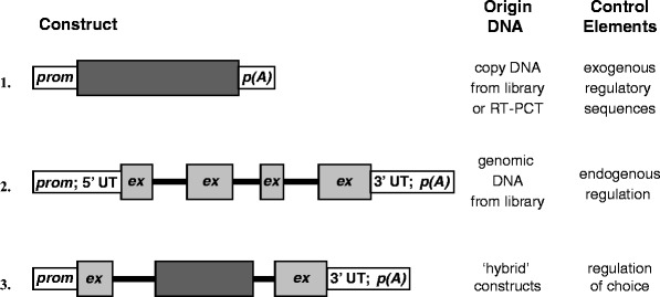
Schematic overview of basic construct design. The basis of the above scheme is the use of eukaryotic coding sequences. The origin of the coding sequences is indicated in the figure. Since regulatory regions are usually not cloned along with cDNA, these have to be provided “separately” and are most often not endogenous (1). Endogenous regulatory elements may be included when the DNA originated from a genomic clone (2). The experimenter has a certain degree of freedom to tailor transgene design to specific requirements ((3); see also Fig. 3). The example depicts a transgene constructed in part of genomic and cDNA sequences. Choice of cDNA and or genomic DNA-based transgenes is discussed in Subheading 1.2. Prom promoter sequences, ex exon, p(A) poly(A+) signal, 5′ and 3′ UT 5′ and 3′ untranslated regions, the thin black lines represent introns (2 and 3).
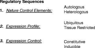
Regulatory sequences in transgene design. Depending on the nature of the animal model and its specific application, there are numerous choices in as far as the regulation of transgene expression is concerned; such regulatory control may comprise more than a promoter only (see Subheading 1.3). (1) Eukaryotic regulatory sequences may be derived from the gene of interest, i.e., autologous (see Subheading 1.3) or from a different gene. (2) The required expression profile may be systemic or tissue specific (see Subheadings 1.3 and 1.4); alternatively (over)expression in all tissues may be achieved with more general promoters. (3) Finally, specific animal models or embryonic lethality may dictate the need for an inducible expression system (see Subheading 1.4). Regulatory sequences of viral origin are widely used to drive transgene expression and frequently confer tissue-specific expression characteristics to a transgene.
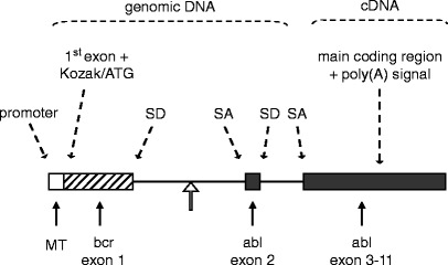
A textbook example of transgene design. The transgene depicted was constructed to study the development of Philadelphia-positive childhood leukemia in a mouse model. The Philadelphia chromosome, the hallmark of this clinical type of leukemia, results from a reciprocal translocation between chromosomes 9 and 22. As a result, the BCR locus and the ABL locus become joined. The genomic ABL locus itself is more than 200 kb in size, the first intron spanning approximately 175 kb. Breakpoints within the ABL locus are known to occur relatively 5′ within the first intron. The transgene harbors a heterologous (see Subheading 1.4) general-type metallothioneine promoter (24). The first BCR exon plus part of the first intron, which is fused (open arrow) to a short part of the first ABL introns and the second ABL exon, provide a splice donor (SD) and a splice acceptor (SA) site and preserve a simple but truthful mammalian intron–exon structure (see Subheading 1.2); an additional SD/SA pair comes from the ABL exon 2/intron 2 and intron 2/exon 3 boundaries. The main body of ABL exons, which spans about 32 kb, was cloned into the transgene as a cDNA segment. In this configuration, the transgene spans a mere 10 kb. Above illustration is adapted after (25).
Similar articles
-
Integrase-Mediated Targeted Transgenics Through Pronuclear Microinjection.Methods Mol Biol. 2020;2066:35-46. doi: 10.1007/978-1-4939-9837-1_3. Methods Mol Biol. 2020. PMID: 31512205
-
Transgene design and delivery into the mouse genome: keys to success.Methods Mol Biol. 2009;561:105-10. doi: 10.1007/978-1-60327-019-9_7. Methods Mol Biol. 2009. PMID: 19504067
-
Generation of transgenic mouse model using PTTG as an oncogene.Methods Mol Biol. 2015;1267:395-411. doi: 10.1007/978-1-4939-2297-0_20. Methods Mol Biol. 2015. PMID: 25636481
-
Insertional mutagenesis in transgenic mice generated by the pronuclear microinjection procedure.Int J Dev Biol. 1998;42(7):1009-17. Int J Dev Biol. 1998. PMID: 9853832 Review.
-
Assembling multiple xenoprotective transgenes in pigs.Xenotransplantation. 2018 Nov;25(6):e12431. doi: 10.1111/xen.12431. Epub 2018 Jul 28. Xenotransplantation. 2018. PMID: 30055014 Review.
Cited by
-
Long terminal repeats power evolution of genes and gene expression programs in mammalian oocytes and zygotes.Genome Res. 2017 Aug;27(8):1384-1394. doi: 10.1101/gr.216150.116. Epub 2017 May 18. Genome Res. 2017. PMID: 28522611 Free PMC article.
References
MeSH terms
LinkOut - more resources
Full Text Sources
