. 2011 Jan;86(1):110-5.
doi: 10.1002/ajh.21888.

A reappraisal of Gaucher disease-diagnosis and disease management algorithms

Affiliations

A reappraisal of Gaucher disease-diagnosis and disease management algorithms

Pramod K Mistry et al. Am J Hematol. 2011 Jan.

Abstract

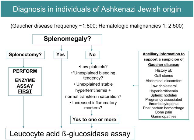
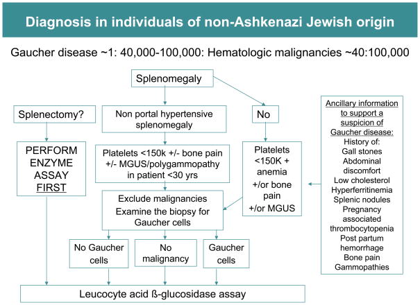
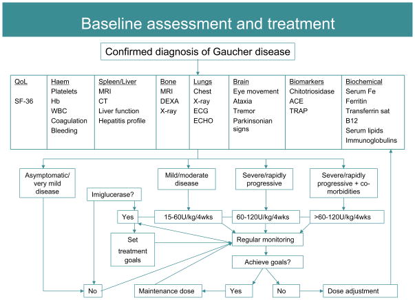
Type 1 (non-neuronopathic) Gaucher disease was the first lysosomal storage disorder for which an effective enzyme replacement therapy was developed and it has become a prototype for treatments for related orphan diseases. There are currently four treatment options available to patients with Gaucher disease, nevertheless, almost 25% of Type 1 Gaucher patients do not gain timely access to therapy because of delays in diagnosis after the onset of symptoms. Diagnosis of Gaucher disease by enzyme testing is unequivocal, but the rarity of the disease and nonspecific and heterogeneous nature of Gaucher disease symptoms may impede consideration of this disease in the differential diagnosis. To help promote timely diagnosis and optimal management of the protean presentations of Gaucher disease, a consensus meeting was convened to develop algorithms for diagnosis and disease management for Gaucher disease.


Figures

FIGURE 1
FIGURE 1
Bone marrow smear and biopsy from a patient with N370S homozygous Gaucher disease showing classical striated macrophages and marrow replacement Smear: Gaucher cells (100×) Core: Bone marrow replacement by Gaucher cells (20×)
FIGURE 1
FIGURE 1
Bone marrow smear and biopsy from a patient with N370S homozygous Gaucher disease showing classical striated macrophages and marrow replacement Smear: Gaucher cells (100×) Core: Bone marrow replacement by Gaucher cells (20×)
FIGURE 2
FIGURE 2
Relationship between age of presentation at extent of splenomegaly (determined by volumetric MRI with normal spleen volume at 0.2% body weight; extent of splenomegaly is indicated by multiple of normal, i.e. xN). Three most common genotypes in North American Gaucher disease patient populations are depicted: N370S/N370S, N370S/L444P and N370S/84G ins G). The results show that the majority of older N370S homozygous patients harbor no, or minimal, splenomegaly)
FIGURE 3
FIGURE 3
(a and b). Comparison of severity of hepatomegaly and bone disease (Herman score [9]) in patients with intact spleen and those with prior splenectomy) stratified for GBA1 gene genotype. The numbers of patients are depicted at the bottom of the bars)
FIGURE 4
FIGURE 4
Diagnosis algorithm for individuals of Ashkenazi origin. Diagnosis of Gaucher disease should be considered in any individual of Ashkenazi Jewish ancestry presenting with mild, moderate or severe splenomegaly. When splenomegaly is absent, Gaucher disease should be considered in the presence of: thrombocytopenia (even if mild), bleeding tendency, unexplained stable hyperferritinemia with normal transferrin saturation, or increased inflammatory markers. *In patients with bleeding diatheses, coagulopathies such as factor XI deficiency common in Askenazim [37] should be excluded.
FIGURE 5
FIGURE 5
Diagnostic algorithm for individuals of non-Ashkenazi Jewish origin. Compared to Gaucher disease, malignancy is likely to be the more frequent cause of splenomegaly in this population. In this setting it is reasonable to perform bone marrow biopsy in initial investigations. The finding of Gaucher cells in bone marrow aspirate will suggest Gaucher disease although Gaucher disease and malignancy are not mutually exclusive. Pseudo-Gaucher cells have also been observed in malignant conditions in the absence of Gaucher disease [38].
FIGURE 6
FIGURE 6
Baseline assessment, treatment and monitoring algorithm. Regular monitoring of patients receiving therapy ensures maximal therapeutic gains through imiglucerase dose adjustment as required. Once therapeutic goals have been met a maintenance dose may be initiated with ongoing monitoring to ensure that disease does not re-establish [50]. Treatment decisions must be personalized; no one treatment regimen will adequately treat all patients.
FIGURE 7
FIGURE 7
(a and b). MRI showing extensive avascular osteonecrosis in the left femur in a previously asymptomatic woman homozygous for N370S mutation with minimal hepatosplenomegaly and cytopenia (b). There is extensive medullary infarction in the left hip that eventually necessitated hip replacement. There is expansion of cellular marrow in the right femur due to marrow infiltration. For comparison, normal marrow MRI appearance is depicted in (a) that shows fatty marrow.
FIGURE 8
FIGURE 8
Liver biopsy showing accumulation of mass of lipid-laden Kupfer cells (hepatic Gaucher cells) in the sinusoids adjacent to normal hepatocytes.

Similar articles

Cited by

References

    1. Beutler E, Grabowski GA. Gaucher disease. In: Scriver CR, Beaudet AL, Sly WS, Valle D, editors. The metabolic and molecular basis of inherited disease. New York, NY: McGraw-Hill; 2001. pp. 3635–3668.
    1. Zimran A, Altarescu G, Rudensky B, et al. Survey of hematological aspects of Gaucher disease. Hematology. 2005;10:151–156. - PubMed
    1. Cox TM, Schofield JP. Gaucher’s disease: clinical features and natural history. Baillieres Clin Haematol. 1997;10:657–89. - PubMed
    1. Grabowski GA. Gaucher disease: gene frequencies and genotype/phenotype correlations. Genet Test. 1997;1:5–12. - PubMed
    1. Horowitz M, Pasmanik-Chor M, Borochowitz Z, et al. Prevalence of glucocerebrosidase mutations in the Israeli Ashkenazi Jewish population. Hum Mutat. 1998;12:240–244. - PubMed

Publication types