. 2010 Nov 16:11:86.
doi: 10.1186/1471-2199-11-86.

Germline transformation of the stalk-eyed fly, Teleopsis dalmanni

Affiliations

Germline transformation of the stalk-eyed fly, Teleopsis dalmanni

Ian A Warren et al. BMC Mol Biol. .

Abstract

Background: Stalk-eyed flies of the family Diopsidae have proven to be an excellent model organism for studying the evolution of ornamental sexual traits. In diopsid flies the eyes and antennae are borne at the end of lateral head projections called 'eye-stalks'. Eyespan, the distance between the eyes, and the degree of sexual dimorphism in eyespan vary considerably between species and several sexually dimorphic species show sexual selection through female mate preference for males with exaggerated eyespan. Relatively little is known about the molecular genetic basis of intra- or inter-species variation in eyespan, eye-stalk development or growth regulation in diopsids. Molecular approaches including comparative developmental analyses, EST screening and QTL mapping have identified potential candidate loci for eyespan regulation in the model species Teleopsis dalmanni. Functional analyses of these genes to confirm and fully characterise their roles in eye-stalk growth require the development of techniques such as germline transformation to manipulate gene activity in vivo.

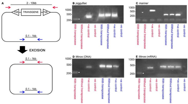
Results: We used in vivo excision assays to identify transposon vector systems with the activity required to mediate transgenesis in T. dalmanni. Mariner based vectors showed no detectable excision while both Minos and piggyBac were active in stalk-eyed fly embryos. Germline transformation with an overall efficiency of 4% was achieved using a Minos based vector and the 3xP3-EGFP marker construct. Chromosomal insertion of constructs was confirmed by Southern blot analysis. Both autosomal and X-linked inserts were recovered. A homozygous stock, established from one of the X-linked inserts, has maintained stable expression for eight generations.

Conclusions: We have performed stable germline transformation of a stalk-eyed fly, T. dalmanni. This is the first transgenic protocol to be developed in an insect species that exhibits an exaggerated male sexual trait. Transgenesis will enable the development of a range of techniques for analysing gene function in this species and so provide insight into the mechanisms underlying the development of a morphological trait subject to sexual selection. Our X-linked insertion line will permit the sex of live larvae to be determined. This will greatly facilitate the identification of genes which are differentially expressed during eye-stalk development in males and females.


Figures

Figure 1
Figure 1
PCR-based assays for transposon excision in T. dalmanni embryos. (A) Schematic representation of a PCR-based excision assay. Donor plasmids contain the terminal inverted repeats (TIRs), from the piggyBac, mariner or Minos transposon, flanking a transgene. In the presence of transposase, donor plasmids undergo excision of the transgene. "Excision primers" (red arrows) flank the entire construct including the TIRs. PCR of the unexcised construct will give rise to a product too large to be amplified efficiently (2-10 kb) under standard conditions. Amplification post-excision produces a smaller product (0.1-1 kb). Excision primers are validated using modified donor plasmids, from which the element had been excised by digestion with an appropriate restriction enzyme, as control templates. "Extraction primers" (blue arrows) amplify part of the donor plasmid backbone to demonstrate successful extraction of the donor plasmid from injected embryos. PCR with these primers produces the same size product (0.1-1 kb) both pre-and post-excision. (B-E) Results of the excision assay using piggyBac (B), mariner (C) and Minos (D and E). Templates for PCR were: DNA extracted from embryos injected with donor plasmid and a source of transposase (with transposase); DNA extracted from embryos with donor plasmid without a source of transposase (without transposase); donor plasmid control templates (+ve control); water (-ve control). PCR reactions either used excision primers (red lettering) to test for excision of the transposable element, or extraction primers (blue lettering) to test for successful extraction of the plasmids from injected embryos (see Additional file 2: Table S1). White triangles denote the expected size of the excision primer PCR product post-excision of the element. For the piggyBac (B) and mariner (C) assays, a DNA source of transposase was used. For the Minos assays both a DNA source of transposase (D) and an mRNA source of transposase were tested (E). Excision was detected for piggyBac and both Minos assays when the donor plasmid was injected with a source of transposase but not in the mariner assay. No excision was detected when donor plasmids were injected without a source of transposase indicating a lack of endogenous transposase activity in the embryos. In all cases donor plasmids were successfully extracted from embryos.
Figure 2
Figure 2
EGFP expression in transgenic larvae. Larvae are from line 34.6 (A, B), line 17.1 (D,E) or non-transgenic (C). In both transgenic lines EGFP fluorescence was restricted to the posterior-most segments of the larva. Asterisks denote anal pads and ps denotes posterior spiracles. (A) Posterior end of a first instar larva showing EGFP expression in the anal pads. Note the position of the posterior spiracles. (B) Posterior end of a third instar larva showing strong EGFP fluorescence in mature anal pads. (C) Posterior end of a non-transgenic third instar larva. A degree of autofluorescence is visible in the cuticle but not in the anal pads. (D) Full length third instar larva showing EGFP expression at the posterior end. Autofluorescence is visible in the gut. (E) High power view of anal pad EGFP expression of the larva shown in (D). Scale bar in A = 100 μm; in B, C, and E = 200 μm; in D = 1 mm.
Figure 3
Figure 3
Molecular characterisation of insertions. (A). Southern analysis confirms transgene integration. DNA was extracted from individuals from four G1 individual derived lines carrying the transgene (17.1, 17.2, 34.6 & 34.14) and digested with EcoRI. The probe was generated from the NotI/SalI fragment of pMi[3xP3-EGFP]. DNA from wild-type (WT) individuals was included as a control. For lines 17.1 and 17.2 single bands of the same size (approximately 6500 bp) are present. For lines 34.6 and 34.14 the multiple bands are indicative of multiple inserts at different locations in the genome. No bands are present in wild-type DNA. (B). Flanking sequence of right arm of an insert present in subline 34.6. The pMi[3xP3-EGFP] sequence is shown in upper case and the flanking genomic sequence in lower case. TA duplication, characteristic of a Minos mediated insertion event, is underlined.

Similar articles

Cited by

References

    1. Darwin C. The Descent of Man, and Selection in Relation to Sex. London: Murray; 1871.
    1. Andersson M. Sexual Selection. New Jersey: Princeton University Press; 1994.
    1. Wilkinson GS. In: Model Systems in Behavioural Ecology. Integrating Conceptual, Theoretical, and Empirical Approaches. Dugatkin LA, editor. Princeton: Princeton University Press; 2001. Genetic consequences of sexual selection in stalk-eyed flies; pp. 72–91.
    1. Chapman T, Pomiankowski A, Fowler K. Stalk-eyed flies. Curr Biol. 2005;15:533–535. doi: 10.1016/j.cub.2005.07.015. - DOI - PubMed
    1. Warren I, Smith H. Stalk-eyed flies (Diopsidae): modelling the evolution and development of an exaggerated sexual trait. BioEssays. 2007;29:300–307. doi: 10.1002/bies.20543. - DOI - PubMed

Publication types

Substances