. 2010 Nov 17;99(10):3255-63.
doi: 10.1016/j.bpj.2010.09.028.

Perfringolysin O association with ordered lipid domains: implications for transmembrane protein raft affinity

Affiliations

Perfringolysin O association with ordered lipid domains: implications for transmembrane protein raft affinity

Lindsay D Nelson et al. Biophys J. .

Abstract

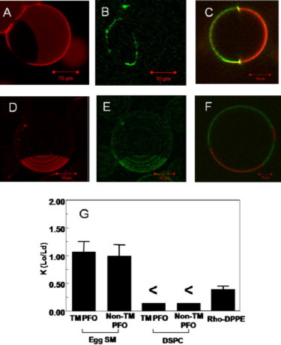
Upon interaction with cholesterol, perfringolysin O (PFO) inserts into membranes and forms a rigid transmembrane (TM) β-barrel. PFO is believed to interact with liquid ordered lipid domains (lipid rafts). Because the origin of TM protein affinity for rafts is poorly understood, we investigated PFO raft affinity in vesicles having coexisting ordered and disordered lipid domains. Fluorescence resonance energy transfer (FRET) from PFO Trp to domain-localized acceptors indicated that PFO generally has a raft affinity between that of LW peptide (low raft affinity) and cholera toxin B (high raft affinity) in vesicles containing ordered domains rich in brain sphingomyelin or distearoylphosphatidylcholine. FRET also showed that ceramide, which increases exposure of cholesterol to water and thus displaces it from rafts, does not displace PFO from ordered domains. This can be explained by shielding of PFO-bound cholesterol from water. Finally, FRET showed that PFO affinity for ordered domains was higher in its non-TM (prepore) form than in its TM form, demonstrating that the TM portion of PFO interacts unfavorably with rafts. Microscopy studies in giant unilamellar vesicles confirmed that PFO exhibits intermediate raft affinity, and showed that TM PFO (but not non-TM PFO) concentrated at the edges of liquid ordered domains. These studies suggest that a combination of binding to raft-associating molecules and having a rigid TM structure that is unable to pack well in a highly ordered lipid environment can control TM protein domain localization. To accommodate these constraints, raft-associated TM proteins in cells may tend to locate within liquid disordered shells encapsulated within ordered domains.


Figures

Figure 1
Figure 1
Sucrose gradient fractionation of TM PFO after treatment of vesicles with TX-100. (A) SM/DOPC/CHOL. (B) SM/CER/DOPC/CHOL. (C) DSPC/DOPC/CHOL. (D) DSPC/DMoPC/CHOL. Both PFO (solid squares, solid line) and control samples (open squares, dashed line) contained 3 μg PFO, a fraction of which was labeled with BODIPY. PFO samples were incubated at room temperature (23°C) with ternary lipid mixtures before solubilization at room temperature. Control samples were incubated at room temperature with binary lipid mixtures DOPC/CHOL (A–C) or DMoPC/CHOL (D), solubilized with TX-100, and then ternary lipid mixtures were added post-solubilization. The Y axis shows the percentage of total BODIPY fluorescence in each fraction.
Figure 2
Figure 2
Fluorescence imaging of TM and non-TM PFO binding to GUVs. (A and B) Three-dimensional reconstructions of a typical 1:1 egg SM/DMoPC with 37% cholesterol GUV after TM PFO binding. The red channel (A) shows the fluorescence signal originating from the Ld marker Rhodamine-DPPE (Rh-DPPE). The dark portion of the bilayer is a Lo domain largely devoid of Rh-DPPE. The green channel (B) shows the signal from BODIPY-labeled TM PFO, which largely accumulates at the Ld/Lo boundary. (C) Confocal image of the equatorial plane of a typical eSM-containing GUV after TM PFO binding. (D and E) Three-dimensional reconstruction of a typical eSM-containing GUV after non-TM PFO binding. The red channel (D) shows Rh-DPPE, and the green channel (B) shows labeled non-TM PFO. (F) Confocal image of the equatorial plane of a typical eSM-containing GUV after non-TM PFO binding. Note that the domains containing the most PFO can vary, and that background intensities have been adjusted to enhance contrast between domains. All scale bars are 10 μm. (G) Protein partition calculated from image intensity analysis (see Materials and Methods) for fluorescent TM PFO and non-TM PFO in both eSM and DSPC-containing GUVS. The value for Rh-DPPE in eSM-containing GUVs is also reported as reference. Error bars show standard deviation (TM PFO in eSM-containing GUVs n = 21, non-TM PFO in eSM-containing GUVs n = 51, TM PFO in DSPC-containing GUVs n = 5, non-TM PFO in DSPC GUVs n = 5, Rh-DPPE n = 27). K(Lo/Ld) equals the ratio of fluorescence intensity in Lo domain divided by that in Ld domains, not counting protein at domain boundaries. The < symbol means that the actual K-value is below our sensitivity for these specific samples (∼0.15). All microscopy experiments were carried out at room temperature.
Figure 3
Figure 3
Schematic illustration of how TM proteins may associate with rafts. Potential locations of TM proteins in membranes with coexisting raft and nonraft domains: (A) Ld shell embedded in the Lo domain, (B) Lo domain or Lo nanodomain, (C) boundary between Lo and Ld domains, and (D) Ld domain or Ld nanodomain . By microscopy, location in a nanodomain or shell of one type would appear as localization in domains of the opposite type (such as in example A).

Similar articles

Cited by

References

    1. Tweten R.K., Parker M.W., Johnson A.E. The cholesterol-dependent cytolysins. Curr. Top. Microbiol. Immunol. 2001;257:15–33. - PubMed
    1. Giddings K.S., Johnson A.E., Tweten R.K. Redefining cholesterol's role in the mechanism of the cholesterol-dependent cytolysins. Proc. Natl. Acad. Sci. USA. 2003;100:11315–11320. - PMC - PubMed
    1. London E. How principles of domain formation in model membranes may explain ambiguities concerning lipid raft formation in cells. Biochim. Biophys. Acta. 2005;1746:203–220. - PubMed
    1. Ohno-Iwashita Y., Shimada Y., Iwashita S. Perfringolysin O, a cholesterol-binding cytolysin, as a probe for lipid rafts. Anaerobe. 2004;10:125–134. - PubMed
    1. Shimada Y., Maruya M., Ohno-Iwashita Y. The C-terminal domain of perfringolysin O is an essential cholesterol-binding unit targeting to cholesterol-rich microdomains. Eur. J. Biochem. 2002;269:6195–6203. - PubMed

Publication types

MeSH terms

LinkOut - more resources