. 2010 Nov 17;99(10):3402-11.
doi: 10.1016/j.bpj.2010.09.021.

Two latent and two hyperstable polymeric forms of human neuroserpin

Affiliations

Two latent and two hyperstable polymeric forms of human neuroserpin

Stefano Ricagno et al. Biophys J. .

Abstract

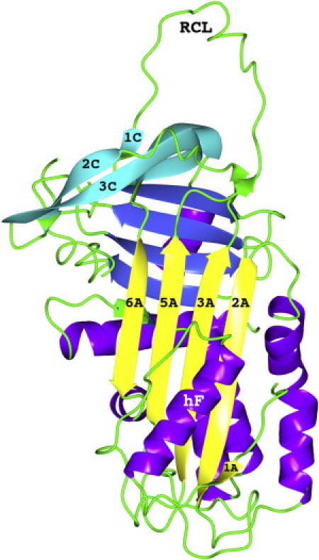
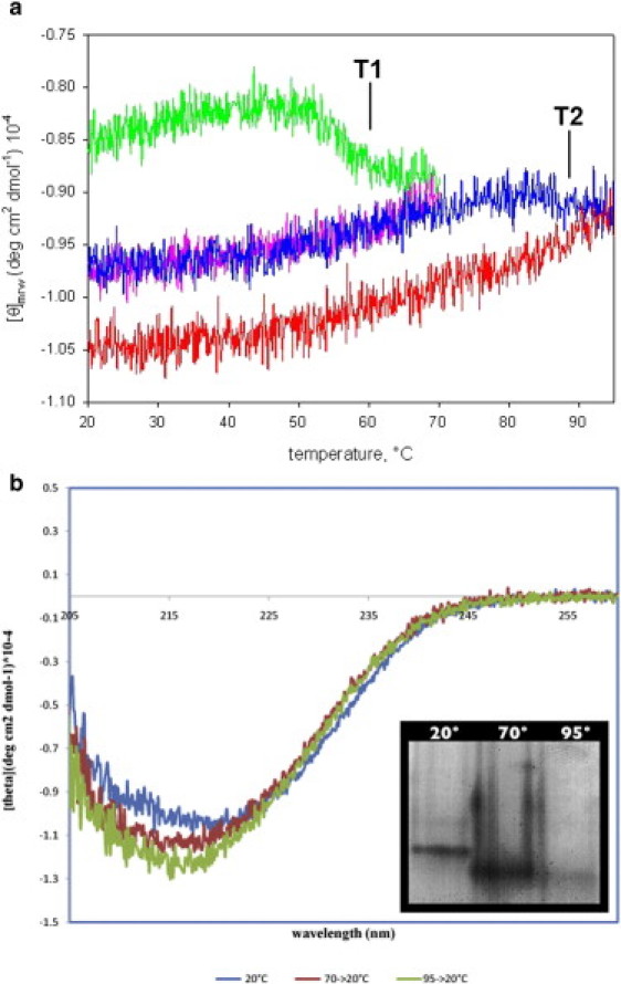
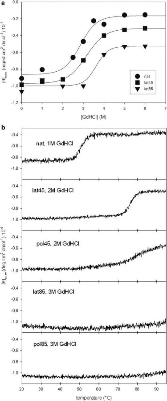
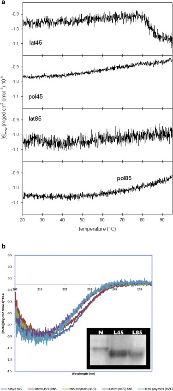
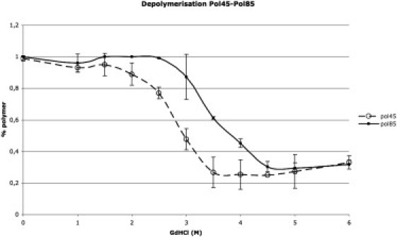
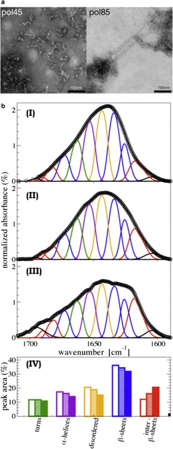
Human neuroserpin (hNS) is a serine protease inhibitor that belongs to the serpin superfamily and is expressed in nervous tissues. The serpin fold is generally characterized by a long exposed loop, termed the reactive center loop, that acts as bait for the target protease. Intramolecular insertion of the reactive center loop into the main serpin β-sheet leads to the serpin latent form. As with other known serpins, hNS pathological mutants have been shown to accumulate as polymers composed of quasi-native protein molecules. Although hNS polymerization has been intensely studied, a general agreement about serpin polymer organization is still lacking. Here we report a biophysical characterization of native hNS that is shown to undergo two distinct conformational transitions, at 55°C and 85°C, both leading to distinct latent and polymeric species. The latent and polymer hNS forms obtained at 45°C and 85°C differ in their chemical and thermal stabilities; furthermore, the hNS polymers also differ in size and morphology. Finally, the 85°C polymer shows a higher content of intermolecular β-sheet interactions than the 45°C polymer. Together, these results suggest a more complex conformational scenario than was previously envisioned, and, in a general context, may help reconcile the current contrasting views on serpin polymerization.


Figures

Figure 1
Figure 1
Cartoon representation of native hNS (pdb code 3F5N). β-Strands belonging to the A- and C-sheets, RCL, and helix F are labeled according to the accepted serpin nomenclature. (A-sheet is shown in yellow, B-sheet in blue, and C-sheet in cyan; helices are purple, and coils, including RCL, are shown in green online).
Figure 2
Figure 2
(a) Temperature ramp of native hNS followed through the far-UV CD signal at 216 nm. In the first segment hNS was heated from 20°C to 70°C (upper trace, green online) and then cooled down to 20°C (middle, purple online). In the third segment hNS was heated to 95°C (middle, blue online) and again cooled down to 20°C (lower trace, red online). The two transitions are highlighted and labeled T1 and T2, respectively. (b) The CD spectra recorded after each step of the temperature ramp are shown at 20°C (upper spectrum, blue online) after being heated at 70°C (middle, red online) and 95°C (lower, green online). (Inset) The same samples analyzed by native PAGE.
Figure 3
Figure 3
(a) Chemical unfolding of native hNS (circles), latent-45 (squares), and latent-85 (triangles), followed through far-UV CD at 216 nm by increasing concentrations of GdHCl (for a more thorough unfolding curve of native hNS and partial unfolding curves for latent forms followed at 222 nm, see Fig. S2). (b) Thermal unfolding (monitored through far-UV CD at 216 nm) of native hNS with 1 M GdHCl, latent-45 with 2 M GdHCl, latent-85 with 3 M GdHCl, polymer-45 with 2 M GdHCl, and polymer-85 with 3 M GdHCl.
Figure 4
Figure 4
(a) Temperature ramps of hNS latent-45, latent-85, polymer-45, and polymer-85 in the 20–95°C temperature range, monitored through far-UV CD at 216 nm. (b) Far-UV CD spectra of native hNS, latent-45, polymer-45, latent-85, and polymer-85 are superimposed (online: native hNS is blue, latent-45 red, polymer-45 green, latent-85 purple, and polymer-85 cyan). (Inset) Native PAGE of native hNS (N), latent-45 (L45), and latent-85 (L85).
Figure 5
Figure 5
Chemical depolymerization of polymer-45 and polymer-85 by increasing concentrations of GdHCl. The black line with triangles and the dashed line with open circles show the polymer-85 and polymer-45 dissolution, respectively. Standard deviations are shown as error bars.
Figure 6
Figure 6
(a) TEM pictures of polymer-45 and polymer-85. (b) FTIR spectra of monomeric native hNS at 25°C (panel I), hNS polymer-45 (panel II), and hNS polymer-85 (panel III). The amide I band areas (1600–1700 cm−1) are normalized to 100 (open circles). Solid lines are the best fit to the data, obtained as the sum of different spectral components (colored curves online). (Panel IV) Percentage of each amide I band spectral component (color-coded online) as for the spectra deconvolution in panels I–III above. Native hNS, open bars; polymer-45, dotted bars; polymer-85, solid bars.

References

    1. Miranda E., Lomas D.A. Neuroserpin: a serpin to think about. Cell. Mol. Life Sci. 2006;63:709–722. - PMC - PubMed
    1. Whisstock J.C., Bottomley S.P. Molecular gymnastics: serpin structure, folding and misfolding. Curr. Opin. Struct. Biol. 2006;16:761–768. - PubMed
    1. Hastings G.A., Coleman T.A., Lawrence D.A. Neuroserpin, a brain-associated inhibitor of tissue plasminogen activator is localized primarily in neurons. Implications for the regulation of motor learning and neuronal survival. J. Biol. Chem. 1997;272:33062–33067. - PubMed
    1. Yepes M., Lawrence D.A. Tissue-type plasminogen activator and neuroserpin: a well-balanced act in the nervous system? Trends Cardiovasc. Med. 2004;14:173–180. - PubMed
    1. Coutelier M., Andries S., Godfraind C. Neuroserpin mutation causes electrical status epilepticus of slow-wave sleep. Neurology. 2008;71:64–66. - PubMed

Publication types