. 2011 Jan 25:1370:16-33.
doi: 10.1016/j.brainres.2010.11.037. Epub 2010 Dec 16.

Multiple neurofilament subunits are present in lamprey CNS

Affiliations

Multiple neurofilament subunits are present in lamprey CNS

Li-Qing Jin et al. Brain Res. .

Abstract

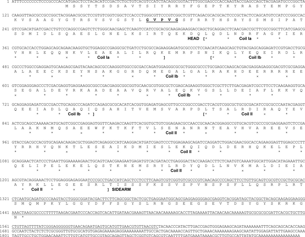
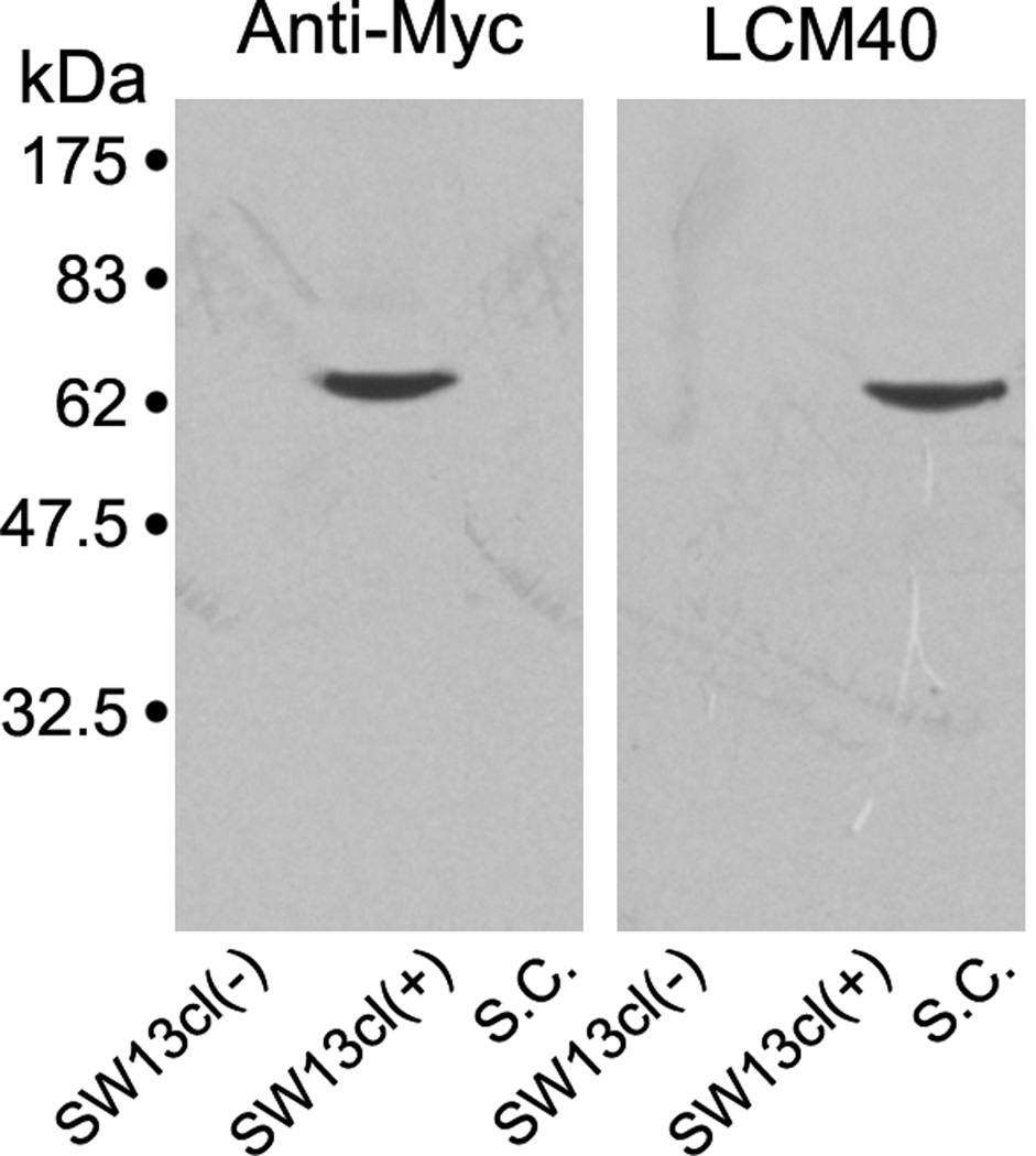
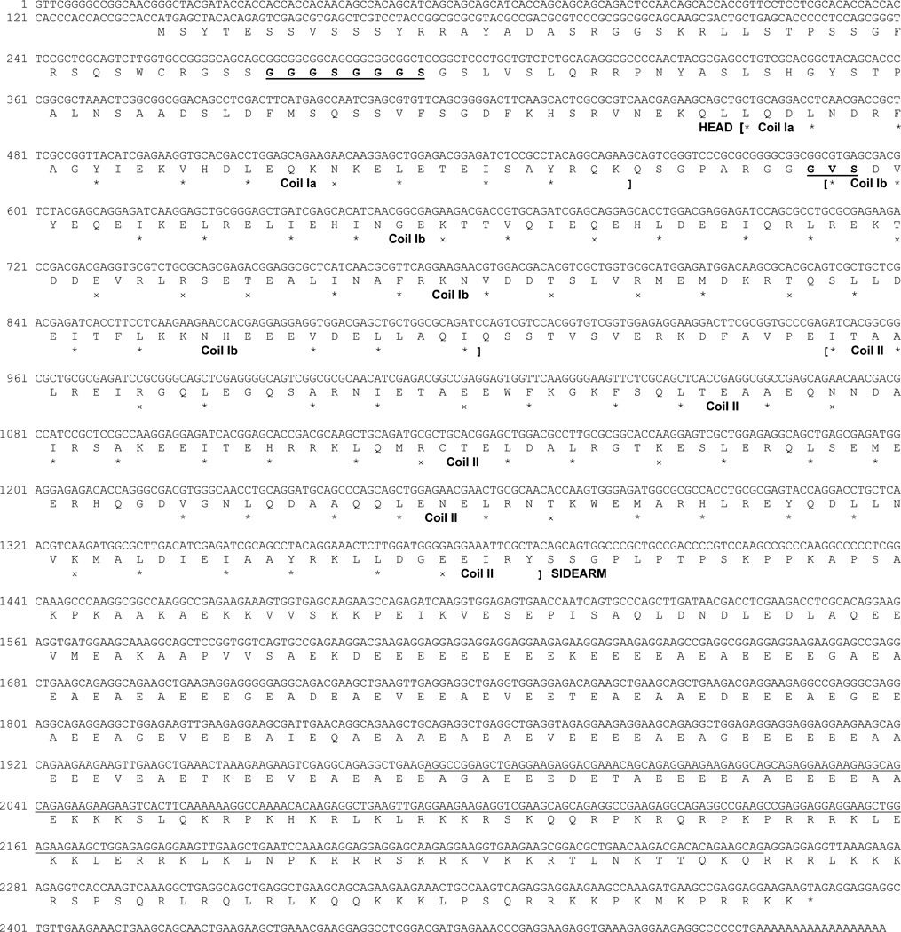
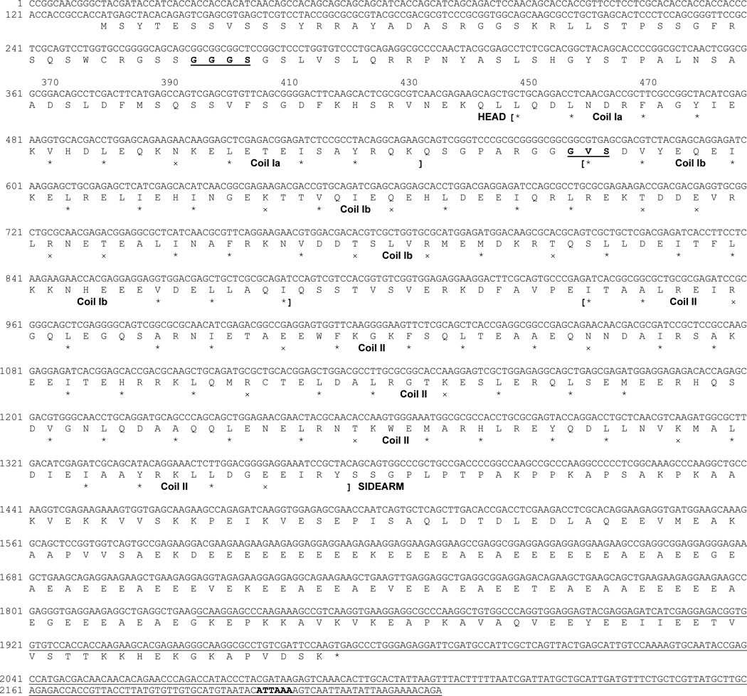
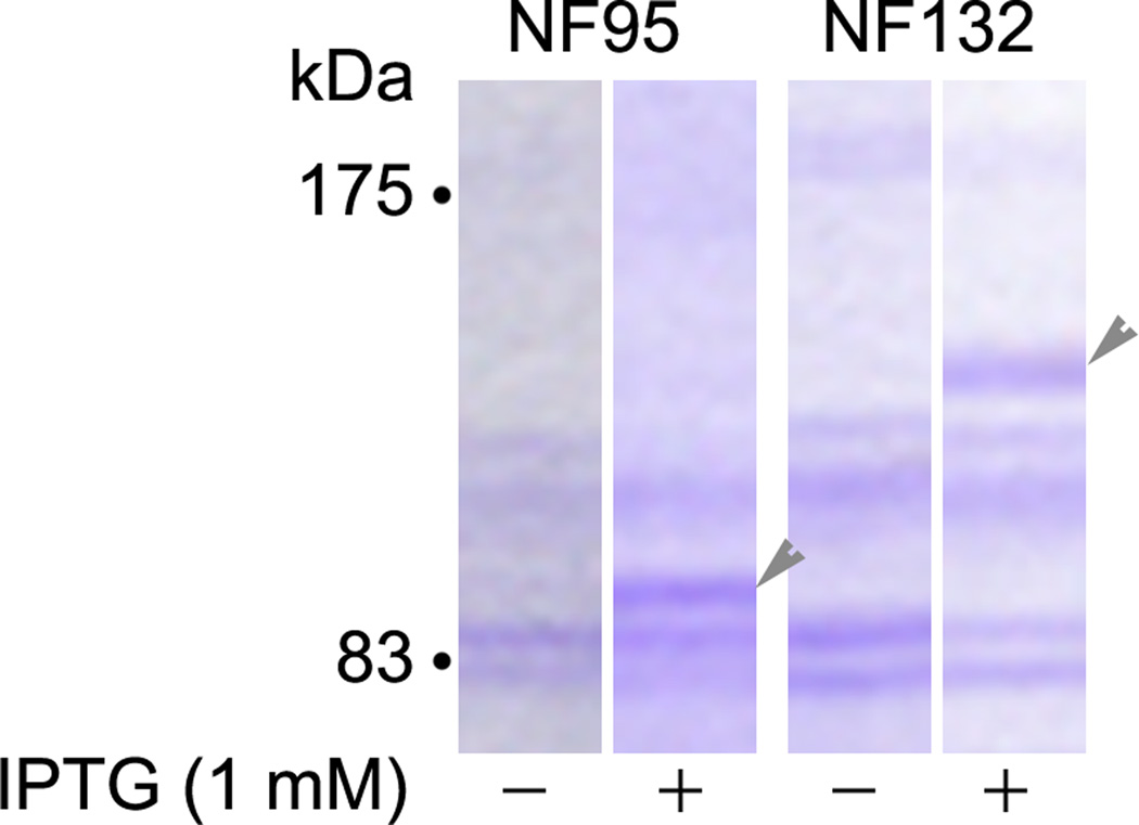
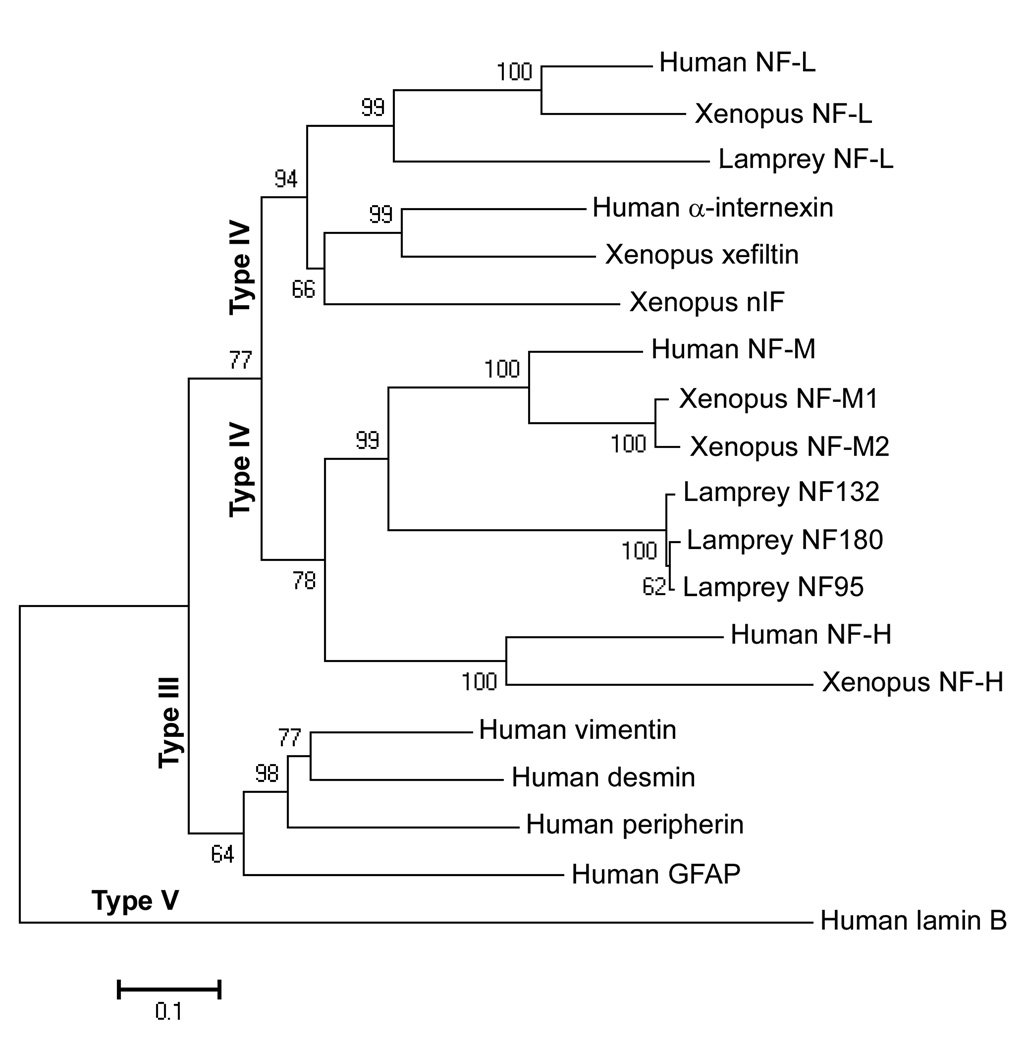
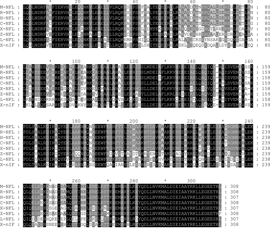
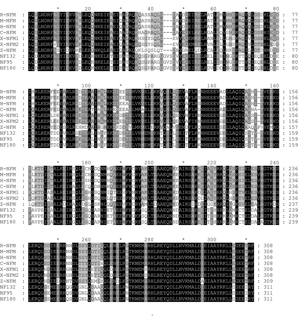
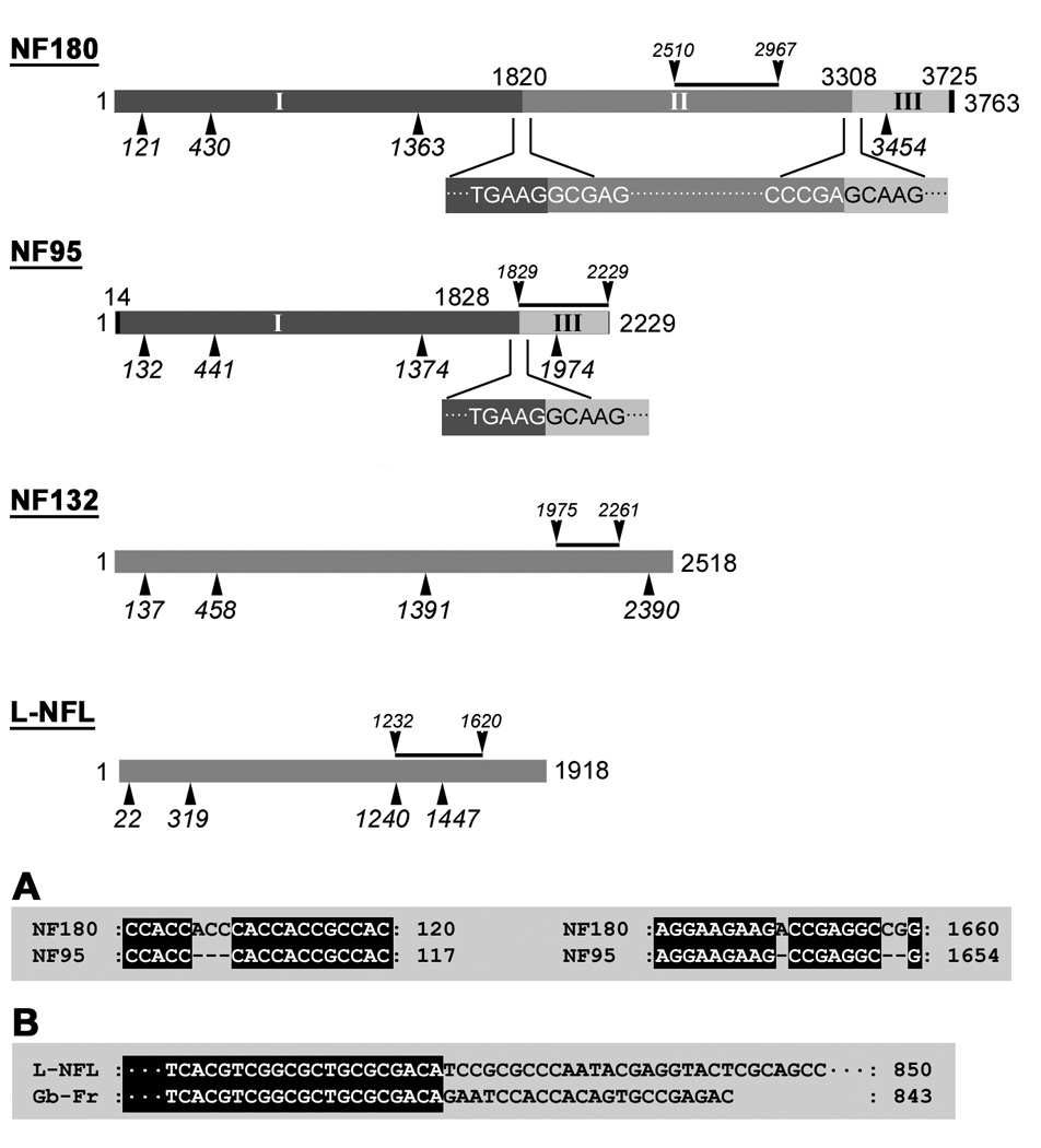
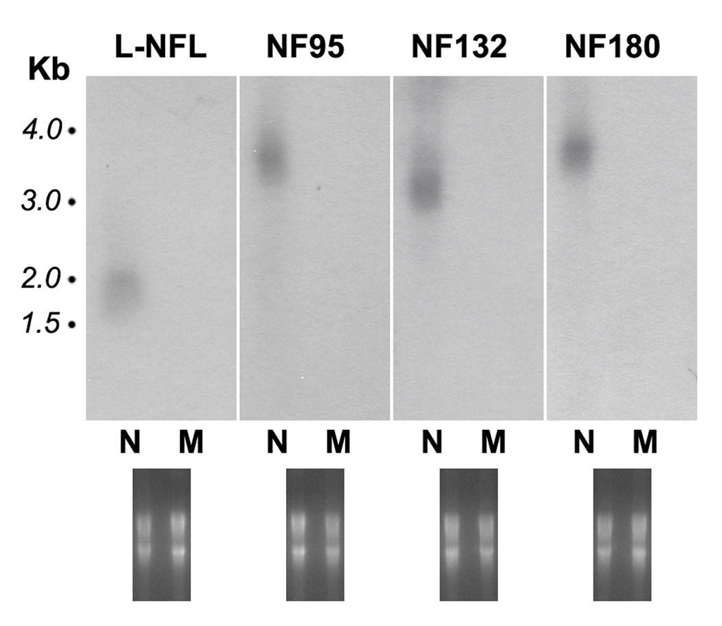
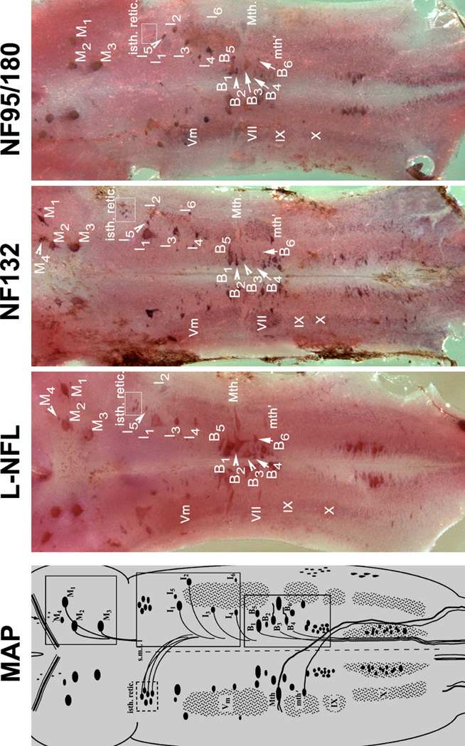
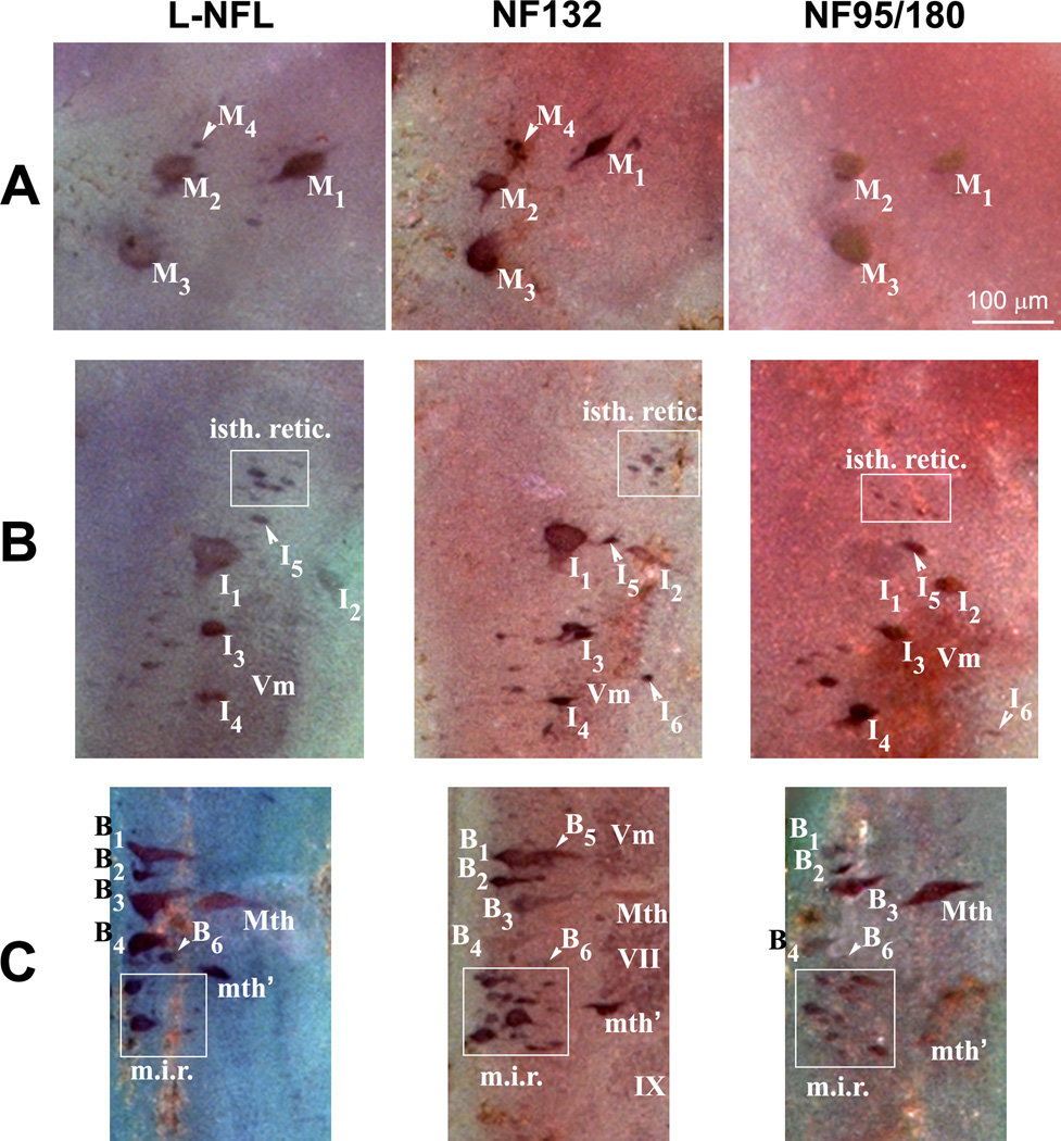
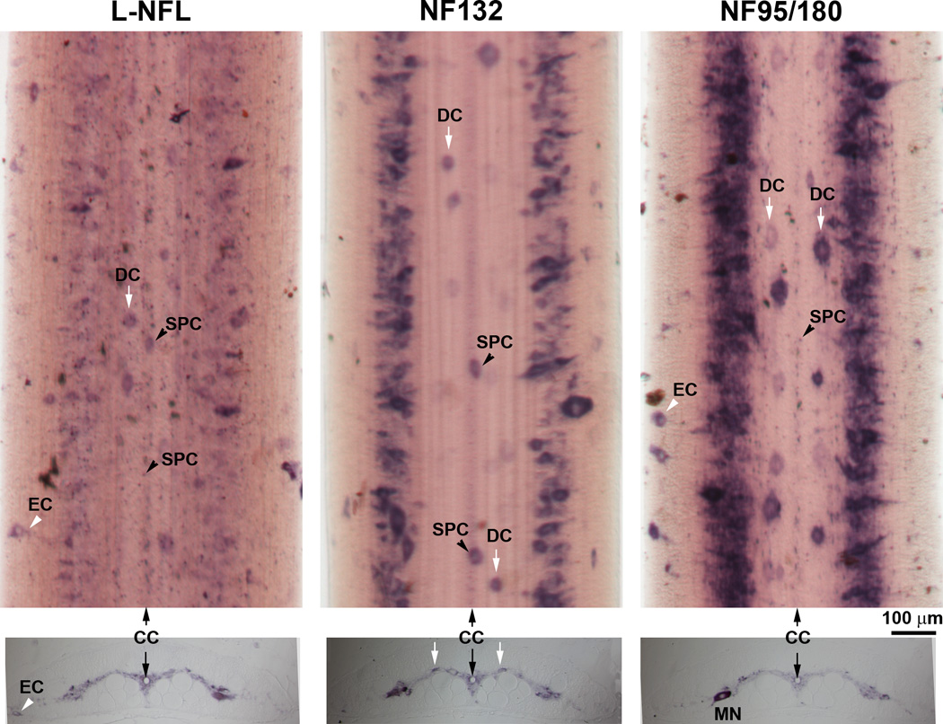
In mammals, there are three neurofilament (NF) subunits (NF-L, NF-M, and NF-H), but it was thought that only a single NF, NF180, exists in lamprey. However, NF180 lacked the ability to self-assemble, suggesting that like mammalian NFs, lamprey NFs are heteropolymers, and that additional NF subunits may exist. The present study provides evidence for the existence of a lamprey NF-L homolog (L-NFL). Genes encoding two new NF-M isoforms (NF132 and NF95) also have been isolated and characterized. With NF180, this makes three NF-M-like isoforms. In situ hybridization showed that all three newly cloned NFs are expressed in spinal cord neurons and in spinal-projecting neurons of the brainstem. Like NF180, there were no KSP multiphosphorylation repeat motifs in the tail regions of NF132 or NF95. NF95 was highly identical to homologous parts of NF180, sharing 2 common pieces of DNA with it. Northern blots suggested that NF95 may be expressed at very low levels in older larvae. The presence of L-NFL in lamprey CNS may support the hypothesis that as in mammals, NFs in lamprey are obligate heteropolymers, in which NF-L is a required subunit.


Figures

Figure 1
Figure 1. Nucleotide and predicted amino acid sequence of the lamprey neurofilament protein L-NFL
Translation was begun at the first in-frame methionine of the longest open reading frame and terminated by the stop codon “TAA”. Nucleotide numbers are indicated at the beginning of each DNA sequence line. The protein sequence before the first bracket is the head region and that after the last bracket is the sidearm (tail) region. Protein sequence between the first and the last bracket is rod region, which consists of 3 coils (coil Ia, Ib, and II). In each coil, stars (*) mark hydrophobic residues at the first and fourth amino acid of putative heptad repeats, while exes (x) mark charged residues. The underlined region is the nucleotide sequence used for Northern blotting or in situ hybridization. The amino acids “GVPVG” in bold and underlined in the head region represents a sequence unique to L-NFL as compared with other NF-Ls listed in Table 1A.
Figure 2
Figure 2. Recombinant L-NFL was not detected by LCM40 in Western blots
The c-Myc-tagged L-NFL recombinant was expressed in SW13cl.2Vim cells. Lysates of the cells with [SW13cl(+)] or without [SW13cl(−)] expression vector were separated on SDS-PAGE with cytoskeletal proteins prepared from lamprey spinal cord (S.C.), and transferred to nitrocellulose membrane. The membranes were then incubated with anti-Myc (Left) or LCM40 (Right) antibody. A band (~ 64 kDa) was labeled by anti-Myc but not by LCM40.
Figure 3
Figure 3. Nucleotide and predicted amino acid sequence of the lamprey neurofilament protein NF132
Translation was begun at the first in-frame methionine of the longest open reading frame and ended by a “TAG” stop codon. Nucleotide numbers are indicated at the beginning of each DNA sequence line. Amino acids “GGGSGGGS” and “GVS” in bold and underlined in the rod region indicates sequences unique to lamprey NF132 as compared with other NF-Ms listed in Table 1B, C.
Figure 4
Figure 4. Nucleotide and predicted amino acid sequence of the lamprey NF protein NF95
Translation was begun at the first in-frame methionine of the longest open reading frame and terminated by a “TGA” stop codon. A potential polyadenylation signal sequence “ATTAAA” in the 3’ unstranslated region is in bold. Note the amino acids “GGGS” and “GVS” in bold and underlined in the rod region. They represent inserted sequences unique to lamprey NF95 and NF180 as compared with other NF-Ms listed in Table 1B, C. Explanations for other marks are the same as in Fig. 1.
Figure 5
Figure 5. Estimation of molecular weights of recombinant NF95 and NF132 in Coomassie Blue-stained SDS gels
The NF132 and NF95 genes were subcloned into the pET-11b (Novagen) expression vector and transfected into BL21(DE3)pLysS cells (Novagen). The cultures were harvested after incubation at 37°C overnight. Expression was induced by IPTG and controls were set without it. The cultures were allowed to grow for another 2 hours and lysed in homogenization buffer. The lysate proteins were separated on a 10% SDS-PAGE, stained with Coomassie Blue R-250 (0.1% in 50% methanol and 10% glacial acetic acid) and destained with 10% methanol and 7% acetic acid. A 95 kDa protein was expressed in NF95-transfected, IPTG-induced cells, while a 132 kDa protein was expressed in NF132-transfected, IPTG-induced cells (arrowheads).
Figure 6
Figure 6. Positions of lamprey NFs in phylogenetic trees constructed with human and Xenopus IFs
Amino acid sequences of 9 human IFs, 6 Xenopus IFs, 1 known lamprey NF (NF180), and 3 putative lamprey NFs (NF132, NF95, and L-NFL) were aligned using the Clustal X tool in BioEdit. The 19 nucleotide sequences obtained from GenBank (accession numbers in parentheses) were: Human desmin (NM_001927), Human GFAP (NM_001131019), Human lamin B (NM_032737), Human NF-H (NM_021076), Human NF-L (NM_006158), Human NF-M (NM_005382), Human peripherin (NM_006262), Human vimentin (NM_003380), Human α-internexin (NM_032727), Lamprey NF132 (DQ398933), Lamprey NF180 (U19361), Lamprey NF95 (DQ398934), Lamprey NF-L (DQ869188), Xenopus NF-H (BC136056), Xenopus NF-L (M86654), Xenopus NF-M1 (U85969), Xenopus NF-M2 (U85970), Xenopus nIF (M86653), Xenopus xefiltin (U63711). Amino acid sequences were inferred from the nucleotide sequences with the ORF finder program of NCBI. A bootstrap test was conducted to construct the NJ tree using MEGA version 4 {Tamura, 2007 #66} (Publication PDF at http://www.-kumarlab.net/publications). Numbers to the left of the branch points indicate the percent of 10,000 bootstrap replicates that support that branch. Lengths in the tree reflect distances between taxa (mean substitutions per residue). Human lamin B was chosen to provide an outgroup for rooting the IF tree because cytoplasmic IF proteins are believed to be derived from an ancestral nuclear lamin {Dodemont, 1990 #17}. Scale: 0.1 amino acid substitutions per site.
Figure 7
Figure 7. Alignment of lamprey NFs with known vertebrate NF sequences
The rod regions of individual NFs were aligned with Clustal X. The generated MSF file was opened and edited with GeneDoc. Conserved amino acid residues are shaded black. Conservative substitutions are in gray. Abbreviations are as in Table 1. A, the amino acid sequence in the rod region of lamprey NF-L was aligned with NF-Ls of other species. B, the amino acid sequences in the rod regions of lamprey NF180, NF132, and NF95 were aligned with NF-Ms of other species. Note the unique sequence “GVS” inserted into the lamprey NF-Ms at position 44–46. At the carboxyl end, a conservative substitution of “D” for “E” occurs at the fourth position of “KLLEGEE” in the lamprey NF-Ms (306th position in the present sequence).
Figure 8
Figure 8. Schematic representations of lamprey NF cDNAs
Four full-length lamprey NF cDNAs are drawn as rods in proportion to their length. The number on the right end represents the nucleotide number. The numbers below the rod represent the numbers of the nucleotides at the beginning of (from left to right) the head, rod, sidearm, and stop codon, respectively. The black line above each rod indicates the region of the individual probe used for Northern blot and in situ hybridizations. Numbers above NF180 and NF95 represent the last nucleotide of the previous fragment. Top 2 rods: NF180 was divided into 3 fragments: I, II, and III. NF95 shared two DNA fragments (I and III) with NF180 but lacked fragment II. Nucleotide sequences between fragments I & II or around fragment II and III in NF180, or the sequences between fragment I & III in NF95, were given below each rod. For fragment I: identity between NF180 and NF95 was 99.0%. For fragment III identity was 99.7%. A: Three gaps in NF95 as compared with NF180 in the fragment I region (ACC106–108, A1650, and CG1658–1659). B: Alignment analysis of L-NFL and an 843 bp sequence of a GenBank DNA fragment (Gb-Fr, TI number: 1229593736) obtained by MegaBlast search of the NCBI trace archive (Petromyzon marinus-WGS).
Figure 9
Figure 9. Northern blot analysis of tissue-specific distributions of lamprey NF mRNAs
Aliquots of 15 µg of total RNA from pooled CNS or muscle from 20 wild-type P. marinus larvae were separated on 1% denaturing agarose gels, capillary blotted to nylon membranes and hybridized with cDNA probes specific to each individual lamprey NF. The blot was exposed to autoradiographic film overnight. Upper panel: For all 4 probes, only a single band was detected, and only in the CNS (N), not in muscle (M). The mRNA sizes for L-NFL, NF95, NF132 and NF180 are ~1.8 kb, 3.7 kb, 3.2 kb, and 3.7 kb. Note: NF95 labeled a band of the same size as NF180. No band showed in the area between L-NFL and NF132 where NF95 mRNA should be. Lower panel: Gels stained with ethidium bromide before transferring to the membrane to show the loading of RNA in each lane.
Figure 10
Figure 10. Expression patterns for lamprey NF mRNAs in the lamprey brain
MAP: Schematic drawing of mature (5-year-old) larval sea lamprey brain stem, showing major anatomical features and the locations of identified neurons and the neuron groups. The view is from the dorsal surface after removal of the mesencephalic and rhombencephalic choroid plexus, transection of the cerebrotectal commissure and obex, and lateral reflection of the alar plate. The identified spinal-projecting neurons include M1-4, I1-6, B1-6, the Mauthner (Mth) and auxiliary Mauthner (Mth’) neurons, the isthmic reticulospinal (isth.retic.), and medial inferior reticulospinal (m.i.r.) cell groups, some cranial nerve nuclei [Vm (trigeminal motor nucleus), VII, IX and X] (Reprinted with permission from Jacobs et al., 1997, J Neurosci 17:5206–5220, ©1997 by the Society for Neuroscience). Three boxes on the right are the locations of 3 identified neuron groups (M-, I-, and B-groups), Mauthner (Mth) and auxiliary Mauthner (mth’) neurons. L-NFL, NF132, and NF95/180 are wholemount in situ hybridizations of lamprey brain stem with corresponding probes. Animals used were large lavae, approximately 4 years old with body lengths 130-, 135-, and 135-mm, respectively. Comparison of regions within boxes are given in Figure 11.
Figure 11
Figure 11. Anatomical distribution of L-NFL, NF132, and NF95/180 in lamprey brain
Based on the nomenclature of Rovainen {Rovainen, 1967 #54} as modified by {Swain, 1993 #64} for identified neurons and neuron groups in mature larval sea lamprey brainstem. M, mesencephalic; I, isthmic; B, bulbar; Mth, Mauthner cell; mth’, auxiliary Mauthner cell. A: Labeled neurons in the mesencephalon. M4 was not detected with the NF95/180 probe. B: Labeled neurons in the isthmic region. Most I group neurons (I1-5), trigeminal nucleus (Vm), and the isthmic reticulospinal neuron group (isth.retic., within white box) were labeled by the probes for L-NFL, NF132, and NF95/180. I6 was not detected with the L-NFL probe. C: Labeled neurons in the middle rhombencephalon. The B1-4 neurons, the Mauthner neuron (Mth), the auxiliary Mauthner neuron (mth’), and the medial inferior reticulospinal (m.i.r., within white box) cell group were labeled by the probes for L-NFL, NF132, and NF95/180. B5 was not detected with the L-NFL and NF95/180 probes.
Figure 12
Figure 12. Cellular localization of NFs in the spinal cord
Upper panel: In situ hybridization in spinal cord wholemounts showed a high level of NF95/180 mRNA expression along the lateral gray matter. L-NFL was expressed relatively evenly and weakly along the entire spinal cord. Several neuron types can be identified by their morphology and location, including: dorsal cells (DC); subependymal cells (SPC) surrounding the central canal (CC), and edge cells (EC). Note: NF132 labeled only large and medium sized cells. Lower panel: In situ hybridization in transverse sections from a 12 cm long larva (approximately 3–4 yrs old). All neurons are localized in the gray matter except for ECs, which sit along the edge of the spinal cord. MN: possible motor neuron, based on size, location and the ventrally projecting axon hillock, which is faintly labeled.

Similar articles

Cited by

References

    1. Asch WS, Leake D, Canger AK, Passini MA, Argenton F, Schechter N. Cloning of zebrafish neurofilament cDNAs for plasticin and gefiltin: increased mRNA expression in ganglion cells after optic nerve injury. J Neurochem. 1998;71:20–32. - PubMed
    1. Bennett GS. Changes in intermediate filament composition during neurogenesis. Curr Top Dev Biol. 1987;21:151–183. - PubMed
    1. Bhat BM, Wold WS. ATTAAA as well as downstream sequences are required for RNA 3'-end formation in the E3 complex transcription unit of adenovirus. Mol Cell Biol. 1985;5:3183–3193. - PMC - PubMed
    1. Breathnach R, Chambon P. Organization and expression of eucaryotic split genes coding for proteins. Annu Rev Biochem. 1981;50:349–383. - PubMed
    1. Bullock TH, Moore JK, Fields RD. Evolution of myelin sheaths: both lamprey and hagfish lack myelin. Neurosci Lett. 1984;48:145–148. - PubMed

Publication types

MeSH terms

Substances