. 2011 Jul;43(1):68-78.
doi: 10.1016/j.nbd.2010.11.003. Epub 2010 Nov 21.

Depletion of Beclin-1 due to proteolytic cleavage by caspases in the Alzheimer's disease brain

Affiliations

Depletion of Beclin-1 due to proteolytic cleavage by caspases in the Alzheimer's disease brain

Troy T Rohn et al. Neurobiol Dis. 2011 Jul.

Abstract

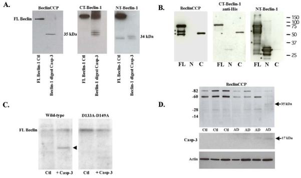
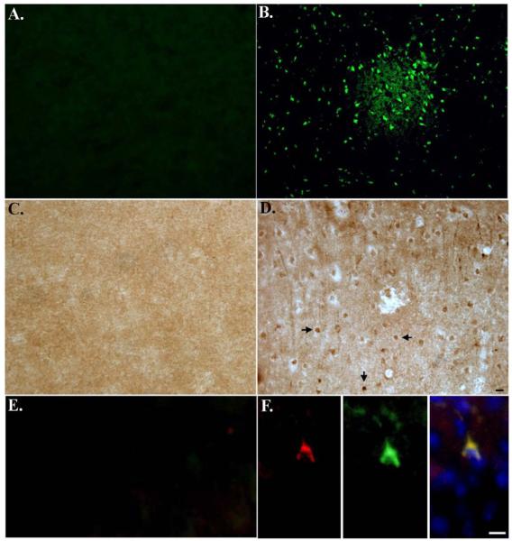
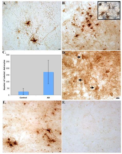
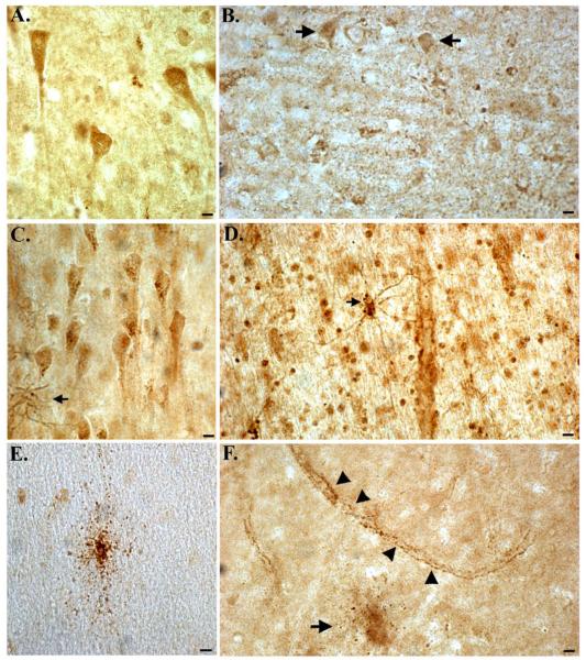
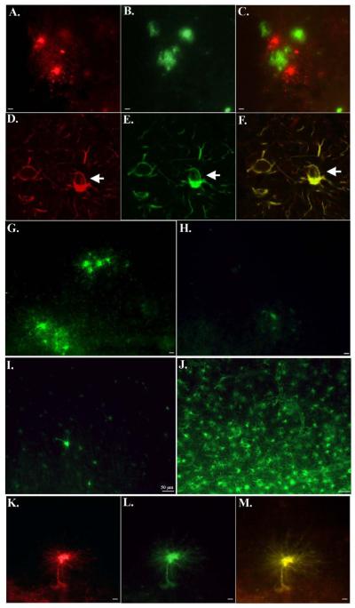
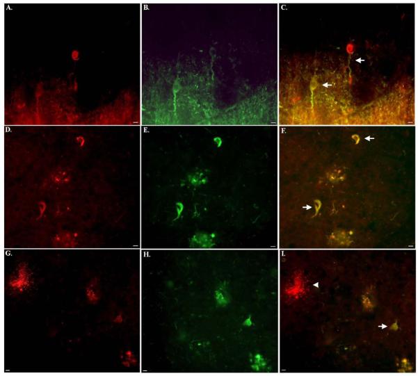
The Beclin-1 protein is essential for the initiation of autophagy, and recent studies suggest this function may be compromised in Alzheimer's disease (AD). In addition, in vitro studies have supported a loss of function of Beclin-1 due to proteolytic modification by caspases. In the present study, we examined whether caspase-cleavage of Beclin-1 occurs in the AD brain by designing a site-directed caspase-cleavage antibody based upon a known cleavage site within the protein at position D149. We confirmed that Beclin-1 is an excellent substrate for caspase-3 and demonstrates cleavage led to the formation of a 35-kDa C-terminal fragment labeled by our novel antibody following Western blot analysis. Application of this antibody termed Beclin-1 caspase-cleavage product antibody or BeclinCCP in frontal cortex tissue sections revealed strong immunolabeling within astrocytes that localized with plaque regions and along blood vessels in all AD cases examined. In addition, weaker, more variable BeclinCCP labeling was also observed within neurofibrillary tangles that colocalized with the early tau conformational marker, MC-1 as well as the late tangle marker, PHF-1. Collectively, these data support a depletion of Beclin-1 in AD following caspase-cleavage. This article is part of a Special Issue entitled "Autophagy and protein degradation in neurological diseases."


Figures

Figure 1
Figure 1. Characterization of BeclinCCP antibody by Western blot analysis
(A): Cell-free incubation of human Beclin-1 by caspase-3 was performed and Western blot analysis was undertaken using three different antibodies as indicated. Panel A, far left depicts undigested Beclin-1 (lane marked Ctl) and Beclin-1 following digest with caspase-3. The BeclinCCP antibody detected the predicted caspase-cleaved C-terminal fragment at 35 kDa. Commercial antibodies to the C-terminal (middle panel) and N-terminal region of Beclin-1 (far right panel) were also tested to confirm the binding pattern of BeclinCCP (see text for details). (B): Recombinant His-tagged Beclin-1-FL, Beclin-1-N and Beclin-1-C were loaded on SDS-PAGE and immunoblotting was performed using the BeclinCCP, anti-pentahis and anti-NT-Beclin-1 antibodies as indicated. Non-specific bands detected on Western are indicated with *. (C): Wild-type Beclin-1 and Beclin-1-D133A-D149A were in vitro transcribed and translated and then left untreated (lane marked Ctl) or incubated with 300 nM of recombinant caspase-3 (lane marked + Casp-3. Cleavage fragments were analyzed by Western blotting using the BeclinCCP antibody. Mutation of these two caspase-cleavage sites prevented proteolytic cleavage and detection of the C-terminal fragment. (D): Western blot analysis with the BeclinCCP antibody or activated capase-3 in soluble human brain extracts. Bottom panel D represents loading control blot following stripping and reprobing using a rabbit antibody to beta-actin (1:400). Data are representative of three independent experiments.
Figure 2
Figure 2. Characterization of BeclinCCP antibody in an in vivo model of apoptosis
Mice were subjected to 1 hr MCAO, followed by reperfusion for 24 hr. (A and B): Brain sections incubated with Fluoro Jade C revealed labeling of shrunken, damaged neurons in the cortex of MCAO mice in ischemic infarct areas (B), while no staining with Fluoro Jade C was evident on the contralateral control side of the brain (A). (C and D): Identical to Panels A and B except sections were analyzed for caspase-cleaved beclin-1 using the BeclinCCP antibody. In this case, BeclinCCP labeling was evident in ischemic infarct areas (D) and was absent on the contralateral side of the brain (C). (E and F): Double-label immunofluorescence experiments with the BeclinCCP antibody (red) and an antibody to activated caspase-3 (green) indicated co-localization of these two markers within ischemic infarct areas (F) that was absent on the contralateral control side of the brain (E). Blue labeling in Panel F (far right) depicts labeling of nuclei with Hoechst. Scale bars represent 10μm.
Figure 3
Figure 3. Characterization of BeclinCCP antibody in the human AD brain by immunohistochemistry
Data are representative staining from human postmortem frontal cortex brain sections following application of the BeclinCCP antibody. (A-B): Application of the BeclinCCP antibody in representative AD cases indicated strong immunolabeling within astrocytes (A) that were localized within plaque-rich regions (B). The inset in Panel B depicts bright-field double-labeling with the BeclinCCP antibody (brown) and a monoclonal antibody to GFAP (blue) (C): Quantification analysis indicated a significant increase in the number of BeclinCCP-positive astrocytes in AD cases (n=11) versus controls (n=5), (p= 0.003). (D): Labeling of the BeclinCCP antibody within apparent NFTs in a representative AD case (arrows, D). (E and F): Serial AD sections were immunolabeled with purified BeclinCCP (1:200) and labeling of astrocytes was observed (E). In contrast, staining with purified BeclinCCP was prevented after preadsorption with free peptide (F). All scale bars represent 10 μm.
Figure 4
Figure 4. Comparison of labeling between the BeclinCCP antibody and commercially available antibodies to full-length beclin-1
(A and B): Representative staining in an age-matched control case (A) or AD case (B) utilizing a commercially available antibody to the N-terminal region of full-length, human beclin-1 revealed labeling of healthy neurons in frontal cortex tissue sections (A), while staining appeared to be diminished in AD (arrows, B). (C and D): Identical to Panels A and B except a commercial antibody to the C-terminal region of beclin-1 was utilized. In this case, labeling was identified within neurons and reactive astrocytes (arrow, C) and in astrocytes along blood vessels in AD cases (D). (E and F): In the same control case, the BeclinCCP antibody only labeled the occasional damaged astrocyte (E), while in AD cases, labeling of astrocytes (arrow, F) and blood vessels (arrowheads, F), was evident. All scare bars represent 10 μm.
Figure 5
Figure 5. Evidence for caspase-cleaved beclin-1 within astrocytes that co-localizes with caspase-cleaved GFAP and are present within plaque-rich regions in the AD brain
(A-C): Double-label immunofluorescence labeling with BeclinCCP in red (A), anti-Aβ antibody in green (B), and the overlap image of both labels (C). Data support the presence of labeling of astrocytes within plaque regions. (D-F): Double-label confocal immunofluorescence images with BeclinCCP (red, D), full-length GFAP antibody (green, E), and the two images overlapped (F). Arrows denote a vacuole within a BeclinCCP-positive astrocyte. (G-J): Single-label immunofluorescence images depicting the inverse relationship between full-length GFAP and BeclinCCP labeling in AD cases. In cases with strong BeclinCCP immunolabeling (G), there was a corresponding lack of staining with the full-length GFAP antibody (H), whereas cases with weak BeclinCCP staining (I) displayed robust labeling with the full-length antibody (J). (K-M): Co-localization of caspase-cleaved beclin-1 and caspase-cleaved GFAP within astrocytes in the AD brain. Data depict labeling utilizing the BeclinCCP antibody in red (K), an antibody to caspase-cleaved GFAP in green (L), and the two images overlapped (M). All scale bars represent 10 μm except panels I and J, which represent 50 μm.
Figure 6
Figure 6. Caspase-cleaved beclin-1 co-localizes with both early and late tangle markers of the Alzheimer’s brain
(A-C): Double-label immunofluorescence images using the PHF-1 antibody (red, A), BeclinCCP antibody (green, B), and the two images overlapped (C). (D-F): Double-label immunofluorescence images using BeclinCCP antibody (red, D), the early tau conformation marker, MC-1 (green, E) and the two images overlapped (F). Yellow, orange coloring represents areas where markers are overlapping, which in this case was evident (arrows, F). (G-I): Identical to (D-F) showing in this case the lack co-localization within a damaged astrocyte labeled with BeclinCCP antibody (arrowhead, I), whereas co-localization between the two antibodies was evident within an apparent tangle-bearing neuron (arrow, I). All scale bars represent 10 μm.

References

    1. Bian GL, et al. Fluoro-Jade C can specifically stain the degenerative neurons in the substantia nigra of the 1-methyl-4-phenyl-1,2,3,6-tetrahydro pyridine-treated C57BL/6 mice. Brain Res. 2007;1150:55–61. - PubMed
    1. Chidlow G, et al. Evaluation of Fluoro-Jade C as a marker of degenerating neurons in the rat retina and optic nerve. Exp Eye Res. 2009;88:426–37. - PubMed
    1. Chun W, Johnson GV. The role of tau phosphorylation and cleavage in neuronal cell death. Front Biosci. 2007;12:733–56. - PubMed
    1. Crews L, et al. Selective molecular alterations in the autophagy pathway in patients with Lewy body disease and in models of alpha-synucleinopathy. PLoS One. 2010;5:e9313. - PMC - PubMed
    1. Donahue JE, Johanson CE. Apolipoprotein E, amyloid-beta, and blood-brain barrier permeability in Alzheimer disease. J Neuropathol Exp Neurol. 2008;67:261–70. - PubMed

Publication types

MeSH terms