. 2011 May;28(5):1581-91.
doi: 10.1093/molbev/msq305. Epub 2010 Nov 15.

Ancestral and derived protein import pathways in the mitochondrion of Reclinomonas americana

Affiliations

Ancestral and derived protein import pathways in the mitochondrion of Reclinomonas americana

Janette Tong et al. Mol Biol Evol. 2011 May.

Abstract

The evolution of mitochondria from ancestral bacteria required that new protein transport machinery be established. Recent controversy over the evolution of these new molecular machines hinges on the degree to which ancestral bacterial transporters contributed during the establishment of the new protein import pathway. Reclinomonas americana is a unicellular eukaryote with the most gene-rich mitochondrial genome known, and the large collection of membrane proteins encoded on the mitochondrial genome of R. americana includes a bacterial-type SecY protein transporter. Analysis of expressed sequence tags shows R. americana also has components of a mitochondrial protein translocase or "translocase in the inner mitochondrial membrane complex." Along with several other membrane proteins encoded on the mitochondrial genome Cox11, an assembly factor for cytochrome c oxidase retains sequence features suggesting that it is assembled by the SecY complex in R. americana. Despite this, protein import studies show that the RaCox11 protein is suited for import into mitochondria and functional complementation if the gene is transferred into the nucleus of yeast. Reclinomonas americana provides direct evidence that bacterial protein transport pathways were retained, alongside the evolving mitochondrial protein import machinery, shedding new light on the process of mitochondrial evolution.


Figures
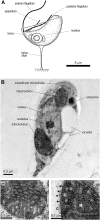
F<sc>IG</sc>. 1.
FIG. 1.
Ultrastructure of R. americana. (A) Drawing of Reclinomonas americana from light micrographs and (B) longitudinal section through cryopreserved samples of R. americana, visualized by TEM. (C) Longitudinal section through posterior mitochondrion to show details of cristae membranes. (D) Higher magnification view of particles (arrows) closely apposed to patches of the mitochondrial outer membrane (M, mitochondrial matrix; C, cytosol).
F<sc>IG</sc>. 2.
FIG. 2.
The mitochondrially-encoded proteome of Reclinomonas americana. (A) Sequence alignment of the mitochondrial RaSecY with SecY from the bacteria Aquifex aeolicus (AaSecY; 21% identity), Thermotoga maritima (TmSecY; 12% identity), and Escherichia coli (EcSecY; 17% identity) highlighting those residues having a conservation score of 7.5 or greater (max 11) with the most conserved residues show in darker colors. The secondary structural elements corresponding to the AaSecY crystal structure (PDB code 3DL8 chain G) are shown with gray lines indicating loops and turns and zig-zags indicating helices. Two insertions are indicated by horizontal orange and green lines and a deletion of an interhelix loop indicated by a red line. The conserved bacterial SecY dibasic motif (KK/RR) is indicated by a red star. (B) AaSecY crystal structure (PDB code 3DL8 chain G; Zimmer et al. 2008) is shown in gold. Highlighted in blue are those conserved residues from RaSecY (conservation score of 7.5 or better) from the sequence alignment. The locations of the two insertions in RaSecY are shown in orange and green and the large loop between transmembrane segments 6 and 7, absent in RaSecY, is shown in red. (C) The mitochondrial genome was conceptually translated and proteome subject to hydropathy analysis as previously described (Chan et al. 2006). An arbitrary line is drawn to distinguish the more hydrophilic proteins (gray-shaded sector) from the more hydrophobic. The hydrophobicity values are provided in supplementary table S1 (Supplementary Material online) (membrane proteins) and supplementary table S2 (Supplementary Material online) (nonmembrane proteins). The individual sequences are color coded according to their predicted location in the inner membrane (black) or matrix (blue). Underlined are those sequences for which a bacterial-type signal sequence or signal anchor was detected (see supplementary table S1, Supplementary Material online). Seven ribosomal proteins (rpS12, S13, S14, L14, L18, L32, L34) have 17 scores below 0 and, being so extremely hydrophilic, are not displayed on this set of axes. Similarly, TatA is not displayed having a mesohydrophobicity score of −4.4 (see supplementary table S1, Supplementary Material online).
F<sc>IG</sc>. 3.
FIG. 3.
Functional complementation of Δcox11 yeast mutants by RaCox11 expressed on a gene in the nucleus. (A) The domain structure of the ScCox11, RaCox11, and MTS-RaCox11 constructs borne by yeast expression plasmids. The MTS-RaCox11 fusion is constructed from residues 1 to 85 of ScCox11 and residues 9–182 from RaCox11. Designated with scissors, an MPP cleavage site is predicted after residue 30 ScCox11. TM denotes the predicted transmembrane segment. The gray oval defines the boundaries of the copper-binding CtaG_Cox11 domain (Pfam 04442) in ScCox11 (residues 105–253) and RaCox11 (residues 25–178). (B) Yeast cells were transformed to express the indicated construct and their growth tested in serial dilution experiments. Equal cell numbers were serially diluted onto medium containing glucose or glycerol as a carbon source and incubated at 25 °C. (C) The transformed yeast strains were grown on medium containing glucose and mitochondria isolated from mid-log phase cultures. Samples of mitochondria (100 μg protein) were analyzed by BN–PAGE, the mitochondrial samples were probed with antisera recognizing the subunit Cox4p to detect cytochrome c oxidase, which migrates as a doublet of bands at ∼1,000 kDa and ∼750 kDa, representing the III2:IV2 and III2:IV supercomplexes formed between cytochrome c oxidase (IV) and cytochrome bc1 reductase (III) as previously described (Schägger and Pfeiffer 2000). Duplicate samples of mitochondria were analyzed by SDS–PAGE and immunoblotting with an antiserum recognizing the outer membrane protein Por1, as a control for the amount of mitochondrial membrane proteins in each of the samples. (D) Transformed yeast cells were grown in liquid media until mid-log phase. The cell numbers measured at 20 h of culture are shown from cultures (white bars) containing glucose as a carbon source, (gray bars) containing glycerol as a carbon source, or (black bars) containing glycerol as a carbon source and supplemented with 50 μM BCDS. The data are representative of five independent experiments.
F<sc>IG</sc>. 4.
FIG. 4.
ScCox11 is imported and assembled via the TIM23 pathway. Mitochondria were isolated from either wild-type yeast or from the tom40–97 yeast strain (Gabriel et al. 2003). Mitochondria (50 μg protein) were incubated with [35S]-labeled Su9-DHFR, PiC, or ScCox11 for the indicated time and analyzed by SDS–PAGE. Precursors (p) were processed to intermediate (i) and mature forms as indicated by arrows. (−ΔΨ) refers to reactions pretreated with 0.1 μM valinomycin. A control lane (T) shows 5% of the total [35S]-labeled precursor proteins.
F<sc>IG</sc>. 5.
FIG. 5.
MTS-RaCox11 is imported more efficiently than RaCox11. (A) Mitochondria were isolated from wild-type yeast and aliquots (50-μg mitochondrial protein) were incubated with [35S]-labeled RaCox11, [35S]-labeled MTS-RaCox11, or [35S]-labeled Su9-DHFR. (−ΔΨ) refers to reactions pretreated with 0.1 μM valinomycin. A control lane (T) shows 5% of the total [35S]-labeled proteins. The import reactions were then treated with trypsin and analyzed by SDS–PAGE and phosphorimage analysis. The mature, processed form of MTS-RaCox11, is indicated by an asterisk (*). (B) Mitochondria were isolated from either wild-type yeast or from the tom40–97 yeast strain and incubated with [35S]-labeled MTS-RaCox11 for the indicated time. The precursor (p) and processed forms are indicated with arrows and (−ΔΨ) refers to reactions pretreated with a cocktail of antimycin, valinomycin, and oligomycin. (C) Mitochondria (100-μg mitochondrial protein) were incubated with [35S]-labeled MTS-RaCox11 for eight min and then reisolated by centrifugation. A sample was prepared for SDS–PAGE (T, total) and a second sample resuspended in 0.1 M sodium carbonate (as described in Materials and Methods) and the membrane pellet (P) and supernatant (S) prepared for SDS–PAGE. The SDS–PAGE gels were analyzed by fluorography and by immunoblots analysis using antisera recognizing the β-subunit of the F1F0-ATP synthase (F1β) or Tim23.
F<sc>IG</sc>. 6.
FIG. 6.
A model for mitochondrial protein sorting in Reclinomonas americana. Proteins like RaCox11, encoded on the mitochondrial genome would be “exported” by the SecY complex; the blue panel denotes that this ancestral protein export pathway would have been present in the bacterium that gave rise to mitochondria but has been lost by most eukaryotes. Genes transferred to the nucleus result in proteins, translated in the cytosol, which must be imported via a TOM complex in the outer membrane. For membrane proteins destined for the inner membrane, insertion and assembly can be via either the TIM23 complex or TIM22 complex. Each of these translocase complexes is composed from members of the Tim23/17/22 family of proteins: in some organisms, there is a single TIM complex (Gentle et al. 2007; Schneider et al. 2008). Reclinomonas americana has at least one member of the Tim23/17/22 protein family (indicated in red, the TBestDB accessions are RAL00006644, RAL00001769, and RAL00003686). Polytopic membrane proteins are ferried to the TIM complex by small TIM chaperones, such as Tim9 and Tim10; R. americana has examples of both proteins (RAL00003758 and RAL00003938, respectively). Imported proteins can be processed by MPPα/β and/or Imp2 and, R. americana has proteases of each type: RAL00003642, RAL00006561, RAL00004675, and RAL00001943.

Similar articles

Cited by

References

    1. Adams KL, Palmer JD. Evolution of mitochondrial gene content: gene loss and transfer to the nucleus. Mol Phylogenet Evol. 2003;29:380–395. - PubMed
    1. Alcock F, Clements A, Webb C, Lithgow T. Tinkering inside the organelle. Science. 2010;327:649–650. - PubMed
    1. Andersson SG, Kurland CG. Origins of mitochondria and hydrogenosomes. Curr Opin Microbiol. 1999;2:535–544. - PubMed
    1. Archibald JM, O'Kelly CJ, Doolittle WF. The chaperonin genes of jakobid and jakobid-like flagellates: implications for eukaryotic evolution. Mol Biol Evol. 2002;19:422–431. - PubMed
    1. Banting GS, Glerum DM. Mutational analysis of the Saccharomyces cerevisiae cytochrome c oxidase assembly protein Cox11p. Eukaryot Cell. 2006;5:568–578. - PMC - PubMed

Publication types

MeSH terms

Substances