. 2011 Jan 14;286(2):1486-98.
doi: 10.1074/jbc.M110.155598. Epub 2010 Nov 16.

Phosphorylation of phosphatidate phosphatase regulates its membrane association and physiological functions in Saccharomyces cerevisiae: identification of SER(602), THR(723), AND SER(744) as the sites phosphorylated by CDC28 (CDK1)-encoded cyclin-dependent kinase

Affiliations

Phosphorylation of phosphatidate phosphatase regulates its membrane association and physiological functions in Saccharomyces cerevisiae: identification of SER(602), THR(723), AND SER(744) as the sites phosphorylated by CDC28 (CDK1)-encoded cyclin-dependent kinase

Hyeon-Son Choi et al. J Biol Chem. .

Abstract

The Saccharomyces cerevisiae PAH1-encoded phosphatidate phosphatase (PAP) catalyzes the penultimate step in the synthesis of triacylglycerol and plays a role in the transcriptional regulation of phospholipid synthesis genes. PAP is phosphorylated at multiple Ser and Thr residues and is dephosphorylated for in vivo function by the Nem1p-Spo7p protein phosphatase complex localized in the nuclear/endoplasmic reticulum membrane. In this work, we characterized seven previously identified phosphorylation sites of PAP that are within the Ser/Thr-Pro motif. When expressed on a low copy plasmid, wild type PAP could not complement the pah1Δ mutant in the absence of the Nem1p-Spo7p complex. However, phosphorylation-deficient PAP (PAP-7A) containing alanine substitutions for the seven phosphorylation sites bypassed the requirement of the phosphatase complex and complemented the pah1Δ nem1Δ mutant phenotypes, such as temperature sensitivity, nuclear/endoplasmic reticulum membrane expansion, decreased triacylglycerol synthesis, and derepression of INO1 expression. Subcellular fractionation coupled with immunoblot analysis showed that PAP-7A was highly enriched in the membrane fraction. In fluorescence spectroscopy analysis, the PAP-7A showed tighter association with phospholipid vesicles than wild type PAP. Using site-directed mutagenesis of PAP, we identified Ser(602), Thr(723), and Ser(744), which belong to the seven phosphorylation sites, as the sites phosphorylated by the CDC28 (CDK1)-encoded cyclin-dependent kinase. Compared with the dephosphorylation mimic of the seven phosphorylation sites, alanine substitution for Ser(602), Thr(723), and/or Ser(744) had a partial effect on circumventing the requirement for the Nem1p-Spo7p complex.


Figures
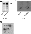
FIGURE 1.
FIGURE 1.
PAP is phosphorylated by yeast and human CDK enzymes. A, purified recombinant PAP (50 μg/ml) was phosphorylated with yeast (1 μg) or human (20 ng) CDK and 20 μm [γ-32P]ATP (2,400 cpm/pmol) for 10 min, followed by SDS-PAGE, transfer to polyvinylidene difluoride membrane, and phosphorimaging analysis. B, polyvinylidene difluoride membranes containing 32P-labeled PAP that were phosphorylated with yeast or human CDK were treated with l-1-tosylamido-2-phenylethyl chloromethyl ketone-trypsin. The resulting peptides were separated on cellulose thin layer plates by electrophoresis (from left to right) in the first dimension and by chromatography (from bottom to top) in the second dimension. The major (labeled 1 and 2) and minor (labeled 3) phosphopeptides were present in the maps of PAP phosphorylated by both CDK enzymes. The radioactive spots labeled 3 in the map for the yeast CDK phosphorylation were observed when the image was overexposed (not shown). C, polyvinylidene difluoride membrane containing 32P-labeled PAP phosphorylated with human CDK was hydrolyzed with 6 n HCl for 90 min at 110 °C, and the hydrolysate was separated by two-dimensional electrophoresis. The positions of phosphorylated PAP and the standard phosphoamino acids phosphoserine (P-Ser), phosphothreonine (P-Thr), and phosphotyrosine (P-Tyr) (dotted lines) are indicated. The data shown are representative of three independent experiments.
FIGURE 2.
FIGURE 2.
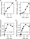
Characterization of CDK activity using PAP as a substrate. Human CDK activity was measured by following the incorporation of the γ-phosphate of [γ-32P]ATP (2,400 cpm/pmol) into purified recombinant PAP under standard phosphorylation conditions except for the variation in time (A), CDK concentration (B), ATP concentration (C), and PAP concentration (D). The CDK reactions were terminated by spotting the mixtures onto P81 phosphocellulose paper. The papers containing the phosphorylated PAP enzyme were washed three times with 75 mm phosphoric acid and then subjected to scintillation counting. The values reported were the average of three experiments ± S.D. (error bars). Some error bars are contained within the symbols.
FIGURE 3.
FIGURE 3.
Effect of CDK on PAP activity. Purified recombinant PAP was phosphorylated with human CDK (20 ng) and 20 μm ATP for the indicated times. Following the phosphorylation reactions, samples were diluted 10-fold, and PAP activity was measured by following the dephosphorylation of 32P-labeled PA. The CDK enzyme was omitted from the control reactions. The values reported were the average of three experiments ± S.D. (error bars). Some error bars are contained within the symbols.
FIGURE 4.
FIGURE 4.
Phosphopeptide mapping analysis of PAP mutants. A, the diagram shows the positions of the NLIP domain, the haloacid dehalogenase (HAD)-like domain, and the serine (S) and threonine (T) residues within a Ser/Thr-Pro motif that were previously identified as sites of phosphorylation (25). B, WT and the indicated phosphorylation site PAP mutant enzymes were expressed and purified from E. coli. The recombinant PAP enzymes were phosphorylated with human CDK (20 ng) and 20 μm [γ-32P]ATP (2,400 cpm/pmol) for 10 min. After phosphorylation, the samples were subjected to SDS-PAGE and transferred to polyvinylidene difluoride membrane. The 32P-labeled proteins were digested with l-1-tosylamido-2-phenylethyl chloromethyl ketone-trypsin. The resulting peptides were separated on cellulose thin layer plates by electrophoresis (from left to right) in the first dimension and by chromatography (from bottom to top) in the second dimension. The radioactive spots labeled 3 were observed when the images were overexposed (not shown). The positions of the phosphopeptides (labeled 1, 2, and 3) that were absent in the S602A, T723A, S744A, and S602A/T723A mutants (indicated by dotted lines) but were present in the wild type enzyme are indicated. The data are representative of three independent experiments.
FIGURE 5.
FIGURE 5.
Effects of the S602A, T723A, and S744A mutations on the time-dependent phosphorylation of PAP. A, WT and the S602A, T723A, and S744A mutant PAP enzymes were expressed and purified from E. coli. The recombinant PAP enzymes (50 μg/ml) were phosphorylated with human CDK (20 ng) and 20 μm [γ-32P]ATP (2,400 cpm/pmol) for the indicated times. After the phosphorylation reactions, the samples were separated by SDS-PAGE; the polyacrylamide gel was dried and then subjected to phosphorimaging analysis. B, the data shown in A were quantified with ImageQuant software. The extent of phosphorylation of the wild type and mutant enzymes at each time point was determined relative to the extent of phosphorylation of the wild type enzyme at 8 min. The maximum amount of phosphorylation for wild type PAP was set at 100%. The data are representative of two independent experiments.
FIGURE 6.
FIGURE 6.
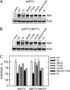
Expression of the phosphorylation-deficient PAP mutant enzymes in S. cerevisiae. Cell extracts were prepared from pah1Δ (A) and pah1Δ nem1Δ (B) cells expressing the indicated WT and PAH1 mutant alleles on low copy plasmids. Samples (40 μg of protein) were subjected to immunoblot analysis using anti-PAP and anti-phosphoglycerate kinase (PGK; loading control) antibodies. C, the relative amounts of PAP/phosphoglycerate kinase proteins from the cells were determined by ImageQuant analysis of the data. Representative immunoblots are shown in A and B, whereas the quantitation data shown in C are the average of three independent experiments ± S.D. (error bars). The positions of the PAP and phosphoglycerate kinase proteins are indicated. 3A, S602A/T723A/S744A triple mutant.
FIGURE 7.
FIGURE 7.
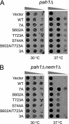
Phosphorylation-deficient PAP complements the temperature-sensitive phenotype of the pah1Δ nem1Δ mutant. Serial dilutions (10-fold) of pah1Δ (A) and pah1Δ nem1Δ (B) cells transformed with low copy pRS415-based plasmids bearing the indicated WT and phosphorylation site mutant forms of PAH1 were spotted onto glucose-containing agar plates. The plates were incubated at the indicated temperatures for 3 days. 3A, S602A/T723A/S744A triple mutant.
FIGURE 8.
FIGURE 8.
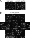
Phosphorylation-deficient PAP-7A complements the nuclear/ER membrane expansion phenotype of the nem1Δ spo7Δ mutant. The nuclear morphology of wild type and nem1Δ spo7Δ mutant cells expressing the HEH2-CHERRY fusion (to label the nucleus) was visualized by fluorescence microscopy. A, wild type and nem1Δ spo7Δ cells expressing the empty vector. B, nem1Δ spo7Δ cells expressing low copy YCplac111-based plasmids bearing the indicated WT and phosphorylation site mutant alleles of PAH1. White bar, 5 μm. 3A, S602A/T723A/S744A triple mutant.
FIGURE 9.
FIGURE 9.
Overexpression of phosphorylation-deficient PAP mutants reduces growth on medium lacking inositol. Serial dilutions (10-fold) of wild type cells transformed with the indicated WT and phosphorylation site mutant forms of PAH1 on galactose (GAL1/10)-inducible high copy YEplac181-based plasmids were spotted on glucose- or galactose-containing agar plates with or without 75 μm inositol and 1 mm choline where indicated. The plates were incubated for 3 days at 30 °C. 3A, S602A/T723A/S744A triple mutant.
FIGURE 10.
FIGURE 10.
Effects of the phosphorylation-deficient PAP mutants on the contents of TAG and phospholipids. pah1Δ (A) and pah1Δ nem1Δ (B) cells expressing the indicated WT and mutant alleles of PAH1 on low copy pRS415-based plasmids were grown to the stationary phase in 5 ml of synthetic complete medium containing [2-14C]acetate (1 μCi/ml). Lipids were extracted and separated by one-dimensional TLC, and the phosphorimages were subjected to ImageQuant analysis. The percentages shown for TAG and phospholipids were normalized to the total 14C-labeled chloroform-soluble fraction. Each data point represents the average of three experiments ± S.D. (error bars). 3A, S602A/T723A/S744A triple mutant.
FIGURE 11.
FIGURE 11.
Effects of the phosphorylation-deficient mutations on the localization of PAP. The indicated WT and phosphorylation-deficient mutant forms of PAP were expressed from a low copy pRS415-based plasmid in pah1Δ (A) and pah1Δ nem1Δ (B) mutant cells. The cells that contained empty vector (V) are also indicated. Extracts (E) prepared from exponential phase cells were fractionated into the cytosolic (C) and membrane (M) fractions by centrifugation. The membrane fraction was resuspended in the same volume as the cytosolic fraction, and equal volumes of the fractions were subjected to immunoblot analysis using anti-PAP, anti-phosphoglycerate kinase (PGK; cytosol marker), and anti-phosphatidylserine synthase (PSS; ER marker) antibodies. C, the relative amounts of cytosol- and membrane-associated PAP were determined for the wild type and mutant forms of the enzyme by ImageQuant analysis of the data. Representative immunoblots are shown in A and B, whereas the quantitation data shown in C are the average of three independent experiments ± S.D. (error bars). The positions of the PAP, phosphoglycerate kinase, and phosphatidylserine synthase (the upper and lower bands are the phosphorylated and dephosphorylated forms of the enzyme, respectively (39)) proteins are indicated. 3A, S602A/T723A/S744A triple mutant.
FIGURE 12.
FIGURE 12.
Effect of the 7A mutations on the interaction of PAP with phospholipid vesicles. A, the indicated WT and phosphorylation-deficient (7A) PAP enzymes that had been expressed and purified from S. cerevisiae were incubated with the indicated concentrations of phospholipid vesicles. Following a 10-min incubation, the increase in PAP fluorescence was measured. The values reported were the average of three experiments ± S.E. (error bars). Some error bars are contained within the symbols. B, dissociation constants (Kd) were determined from the data shown in A.

Similar articles

Cited by

References

    1. Lin Y. P., Carman G. M. (1989) J. Biol. Chem. 264, 8641–8645 - PubMed
    1. Han G. S., Wu W. I., Carman G. M. (2006) J. Biol. Chem. 281, 9210–9218 - PMC - PubMed
    1. Han G. S., Siniossoglou S., Carman G. M. (2007) J. Biol. Chem. 282, 37026–37035 - PubMed
    1. Carman G. M., Han G. S. (2006) Trends Biochem. Sci. 31, 694–699 - PMC - PubMed
    1. Santos-Rosa H., Leung J., Grimsey N., Peak-Chew S., Siniossoglou S. (2005) EMBO J. 24, 1931–1941 - PMC - PubMed

Publication types

MeSH terms