

Molybdopterin dinucleotide biosynthesis in Escherichia coli: identification of amino acid residues of molybdopterin dinucleotide transferases that determine specificity for binding of guanine or cytosine nucleotides
- PMID: 21081498
- PMCID: PMC3020748
- DOI: 10.1074/jbc.M110.155671
Molybdopterin dinucleotide biosynthesis in Escherichia coli: identification of amino acid residues of molybdopterin dinucleotide transferases that determine specificity for binding of guanine or cytosine nucleotides
Abstract
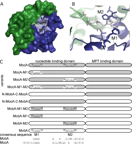
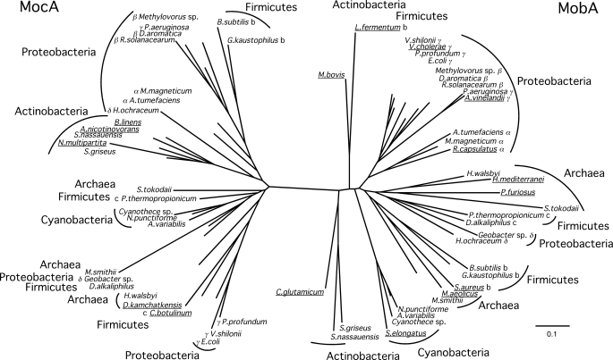
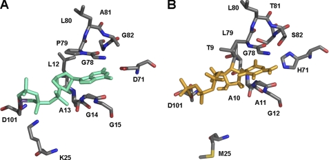
The molybdenum cofactor is modified by the addition of GMP or CMP to the C4' phosphate of molybdopterin forming the molybdopterin guanine dinucleotide or molybdopterin cytosine dinucleotide cofactor, respectively. The two reactions are catalyzed by specific enzymes as follows: the GTP:molybdopterin guanylyltransferase MobA and the CTP:molybdopterin cytidylyltransferase MocA. Both enzymes show 22% amino acid sequence identity and are specific for their respective nucleotides. Crystal structure analysis of MobA revealed two conserved motifs in the N-terminal domain of the protein involved in binding of the guanine base. Based on these motifs, we performed site-directed mutagenesis studies to exchange the amino acids to the sequence found in the paralogue MocA. Using a fully defined in vitro system, we showed that the exchange of five amino acids was enough to obtain activity with both GTP and CTP in either MocA or MobA. Exchange of the complete N-terminal domain of each protein resulted in the total inversion of nucleotide specificity activity, showing that the N-terminal domain determines nucleotide recognition and binding. Analysis of protein-protein interactions showed that the C-terminal domain of either MocA or MobA determines the specific binding to the respective acceptor protein.
Figures
Similar articles
-
MocA is a specific cytidylyltransferase involved in molybdopterin cytosine dinucleotide biosynthesis in Escherichia coli.J Biol Chem. 2009 Aug 14;284(33):21891-21898. doi: 10.1074/jbc.M109.008565. Epub 2009 Jun 19. J Biol Chem. 2009. PMID: 19542235 Free PMC article.
-
The crystal structure of the Escherichia coli MobA protein provides insight into molybdopterin guanine dinucleotide biosynthesis.J Biol Chem. 2000 Dec 22;275(51):40211-7. doi: 10.1074/jbc.M007406200. J Biol Chem. 2000. PMID: 10978347
-
Transfer of the molybdenum cofactor synthesized by Rhodobacter capsulatus MoeA to XdhC and MobA.J Biol Chem. 2007 Sep 28;282(39):28493-28500. doi: 10.1074/jbc.M704020200. Epub 2007 Aug 7. J Biol Chem. 2007. PMID: 17686778
-
The chaperone FdsC for Rhodobacter capsulatus formate dehydrogenase binds the bis-molybdopterin guanine dinucleotide cofactor.FEBS Lett. 2014 Feb 14;588(4):531-7. doi: 10.1016/j.febslet.2013.12.033. Epub 2014 Jan 18. FEBS Lett. 2014. PMID: 24444607
-
The methylator meets the terminator.Proc Natl Acad Sci U S A. 2002 Feb 5;99(3):1104-6. doi: 10.1073/pnas.042004099. Proc Natl Acad Sci U S A. 2002. PMID: 11830650 Free PMC article. Review. No abstract available.
Cited by
-
Structural data on the periplasmic aldehyde oxidoreductase PaoABC from Escherichia coli: SAXS and preliminary X-ray crystallography analysis.Int J Mol Sci. 2014 Jan 31;15(2):2223-36. doi: 10.3390/ijms15022223. Int J Mol Sci. 2014. PMID: 24492481 Free PMC article.
-
A step into the rare biosphere: genomic features of the new genus Terrihalobacillus and the new species Aquibacillus salsiterrae from hypersaline soils.Front Microbiol. 2023 May 9;14:1192059. doi: 10.3389/fmicb.2023.1192059. eCollection 2023. Front Microbiol. 2023. PMID: 37228371 Free PMC article.
-
DMSO Reductase Family: Phylogenetics and Applications of Extremophiles.Int J Mol Sci. 2019 Jul 8;20(13):3349. doi: 10.3390/ijms20133349. Int J Mol Sci. 2019. PMID: 31288391 Free PMC article. Review.
-
The role of FeS clusters for molybdenum cofactor biosynthesis and molybdoenzymes in bacteria.Biochim Biophys Acta. 2015 Jun;1853(6):1335-49. doi: 10.1016/j.bbamcr.2014.09.021. Epub 2014 Sep 28. Biochim Biophys Acta. 2015. PMID: 25268953 Free PMC article. Review.
-
The biosynthesis of the molybdenum cofactors.J Biol Inorg Chem. 2015 Mar;20(2):337-47. doi: 10.1007/s00775-014-1173-y. Epub 2014 Jul 1. J Biol Inorg Chem. 2015. PMID: 24980677
References
-
- Rajagopalan K. V. (1996) in Cellular and Molecular Biology (Neidhardt F. C. ed) pp. 674–679, American Society for Microbiology, Washington, D. C
-
- Rajagopalan K. V., Johnson J. L. (1992) J. Biol. Chem. 267, 10199–10202 - PubMed
-
- Wuebbens M. M., Rajagopalan K. V. (1995) J. Biol. Chem. 270, 1082–1087 - PubMed
-
- Gutzke G., Fischer B., Mendel R. R., Schwarz G. (2001) J. Biol. Chem. 276, 36268–36274 - PubMed
Publication types
MeSH terms
Substances
LinkOut - more resources
Full Text Sources
Molecular Biology Databases
Miscellaneous
