. 2010 Dec;133(Pt 12):3764-77.
doi: 10.1093/brain/awq316. Epub 2010 Nov 16.

Impaired consciousness in temporal lobe seizures: role of cortical slow activity

Affiliations

Impaired consciousness in temporal lobe seizures: role of cortical slow activity

Dario J Englot et al. Brain. 2010 Dec.

Abstract

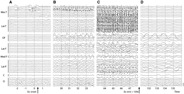
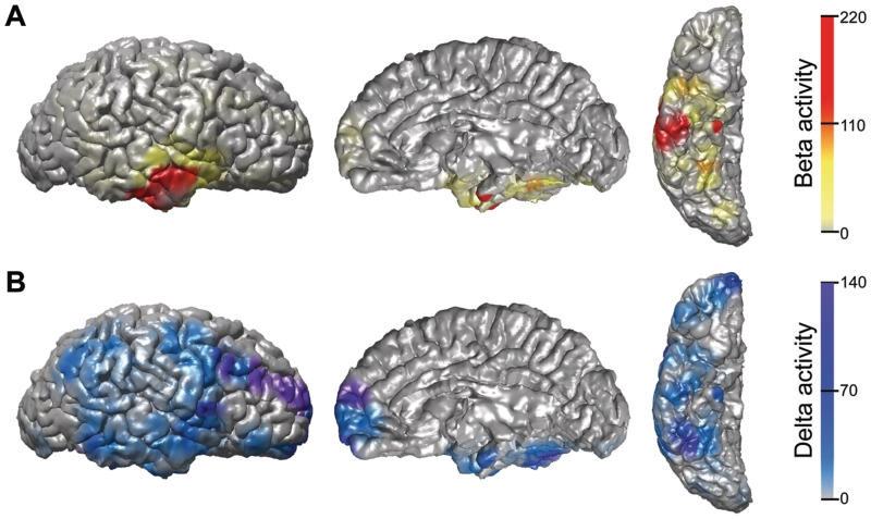
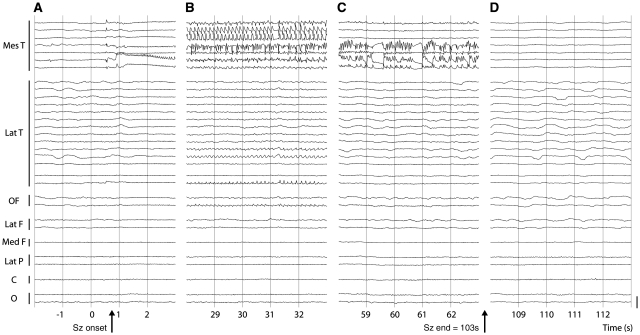
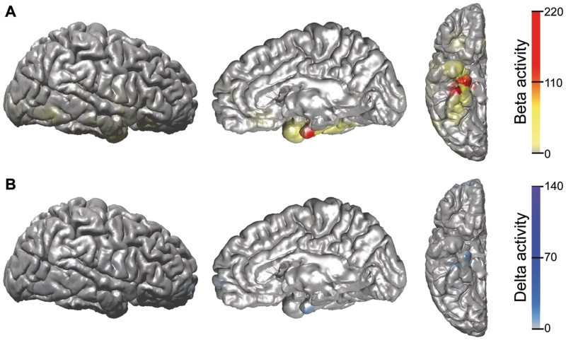
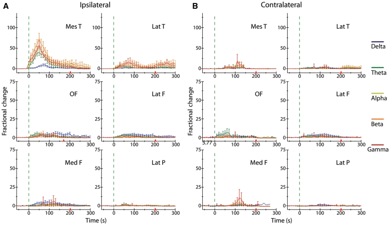
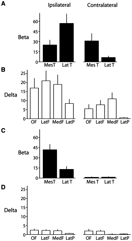
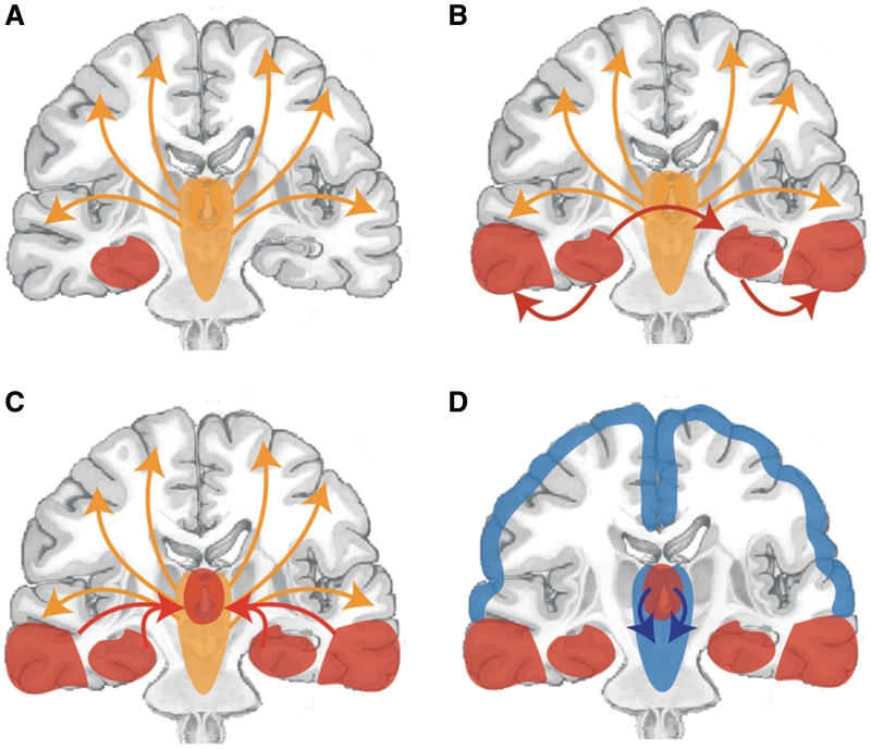
Impaired consciousness requires altered cortical function. This can occur either directly from disorders that impair widespread bilateral regions of the cortex or indirectly through effects on subcortical arousal systems. It has therefore long been puzzling why focal temporal lobe seizures so often impair consciousness. Early work suggested that altered consciousness may occur with bilateral or dominant temporal lobe seizure involvement. However, other bilateral temporal lobe disorders do not impair consciousness. More recent work supports a 'network inhibition hypothesis' in which temporal lobe seizures disrupt brainstem-diencephalic arousal systems, leading indirectly to depressed cortical function and impaired consciousness. Indeed, prior studies show subcortical involvement in temporal lobe seizures and bilateral frontoparietal slow wave activity on intracranial electroencephalography. However, the relationships between frontoparietal slow waves and impaired consciousness and between cortical slowing and fast seizure activity have not been directly investigated. We analysed intracranial electroencephalography recordings during 63 partial seizures in 26 patients with surgically confirmed mesial temporal lobe epilepsy. Behavioural responsiveness was determined based on blinded review of video during seizures and classified as impaired (complex-partial seizures) or unimpaired (simple-partial seizures). We observed significantly increased delta-range 1-2 Hz slow wave activity in the bilateral frontal and parietal neocortices during complex-partial compared with simple-partial seizures. In addition, we confirmed prior work suggesting that propagation of unilateral mesial temporal fast seizure activity to the bilateral temporal lobes was significantly greater in complex-partial than in simple-partial seizures. Interestingly, we found that the signal power of frontoparietal slow wave activity was significantly correlated with the temporal lobe fast seizure activity in each hemisphere. Finally, we observed that complex-partial seizures were somewhat more common with onset in the language-dominant temporal lobe. These findings provide direct evidence for cortical dysfunction in the form of bilateral frontoparietal slow waves associated with impaired consciousness in temporal lobe seizures. We hypothesize that bilateral temporal lobe seizures may exert a powerful inhibitory effect on subcortical arousal systems. Further investigations will be needed to fully determine the role of cortical-subcortical networks in ictal neocortical dysfunction and may reveal treatments to prevent this important negative consequence of temporal lobe epilepsy.


Figures

Figure 1
Figure 1
Example intracranial EEG recording during a temporal lobe complex-partial seizure. (A) Seizure (Sz) onset with low-voltage fast activity emerging from periodic spiking in the mesial temporal contacts. (B) Sample EEG from early seizure. Rhythmic poly-spike and sharp wave activity develops in the mesial temporal lobe, while the frontal and parietal contacts show large-amplitude irregular slow activity. (C) Sample EEG from mid-seizure. Poly-spike and wave activity is present in the mesial and lateral temporal lobe contacts, with ongoing slow waves in the association cortex. Rolandic and occipital contacts are relatively spared. (D) Postictal suppression is seen in temporal lobe contacts, with continued irregular slowing in the frontoparietal neocortex. Only ipsilateral contacts are shown. Bars along left margin indicate electrode contacts from different strips, grids or depth electrodes in the indicated brain regions. A subset of representative electrodes are shown of the 128 studied in this patient. Calibration bar on right is 3 mV. Montage is referential to mastoid. Mes T = mesial temporal; Lat T = lateral temporal; OF = orbital frontal; Lat F = lateral frontal; Med F = medial frontal; Lat P = lateral parietal; C = perirolandic pre- and post-central gyri; O = occipital.
Figure 2
Figure 2
Three dimensional colour maps of beta and delta activity changes during a complex-partial seizure. (A) Large elevations in fast beta activity are seen in the mesial and lateral temporal lobe during the event, representing regions of seizure onset and propagation, respectively. (B) Increased delta activity is most dramatic in the frontal and parietal association cortices, where there is no fast seizure activity (A), as well as in the mesial and lateral temporal lobe, where fast seizure activity is also present (A). The occipital and perirolandic areas are relatively spared. Data shown are fractional change in beta- (A) or delta- (B) range EEG signal power during the entire seizure versus a 60 s uninterrupted baseline, overlaid on lateral, medial and ventral views of a 3D reconstruction of the patient’s pre-implant MRI. Electrode locations are shown in Supplementary Fig. 1A. Only the ipsilateral (right) hemisphere is shown. Raw EEG of the same seizure is shown in Fig. 1 and 3D colour map time course is shown in Supplementary Video 1.
Figure 3
Figure 3
Example intracranial EEG recording during a temporal lobe simple-partial seizure. (A) Seizure (Sz) onset with low-voltage fast activity emerging from periodic spiking in the mesial temporal contacts. (B) Sample EEG from early seizure. Rhythmic poly-spike and sharp wave activity continues in the mesial temporal lobe. Few changes are observed in lateral temporal and other cortical contacts. (C) Sample EEG from mid-seizure. Poly-spike and wave activity continues in the mesial temporal lobe, while other contacts show activity resembling pre-seizure baseline. No large-amplitude cortical slow activity is seen. (D) Postictal suppression is seen in the mesial temporal lobe. Only ipsilateral contacts are shown. Bars along left margin indicate electrode contacts from different strips, grids or depth electrodes in the indicated brain regions. A subset of representative electrodes are shown of the 128 studied in this patient. Calibration bar on right is 3 mV. Montage is referential to mastoid. Mes T = mesial temporal; Lat T = lateral temporal; OF = orbital frontal; Lat F = lateral frontal; Med F = medial frontal; Lat P = lateral parietal; C = perirolandic pre- and post-central gyri; O = occipital.
Figure 4
Figure 4
Three dimensional colour maps of beta and delta activity changes during a simple-partial seizure. (A) Elevations in fast beta activity are seen in the mesial temporal lobe, the site of seizure onset, with minimal lateral temporal involvement. (B) Increased delta activity is seen in the mesial temporal lobe, in the same region as the ictal beta activity, but the neocortex is relatively spared. Data shown are fractional change in beta- (A) or delta- (B) range EEG signal power during the entire seizure versus a 60 s uninterrupted baseline, overlaid on lateral, medial and ventral views of a 3D reconstruction of the patient’s pre-implant MRI. Electrode locations are shown in Supplementary Fig. 1B. Only the ipsilateral (right) hemisphere is shown. Raw EEG of the same seizure is shown in Fig. 3 and 3D colour map time course is shown in Supplementary Video 2.
Figure 5
Figure 5
Time course plots of intracranial EEG changes during complex-partial seizures. (A) Ipsilateral to onset of complex-partial seizures, dramatic increases in fast EEG frequency bands (theta, alpha, beta, gamma) are observed in the mesial and lateral temporal lobe contacts compared with baseline. EEG power in ipsilateral frontoparietal neocortical regions also increases considerably during and following seizures in all frequency bands, with largest increases in slow delta activity in the orbital, lateral and medial frontal cortices. (B) Contralateral to onset of complex-partial seizures, large increases in fast EEG frequency bands (theta, alpha, beta, gamma) are observed in the mesial temporal lobe contacts compared with baseline, with smaller increases in the contralateral lateral temporal lobe. Increases in slow delta EEG activity are recorded in the frontal cortex ictally and postictally. Relatively few changes are observed in the contralateral lateral parietal cortex. Data are mean fractional change (±SEM) in EEG power from 60 s pre-seizure baseline binned every 10 s. Vertical dotted lines indicate seizure onset; arrows indicate mean seizure offset time. Note: different scale between temporal (top) and neocortical (middle and bottom) data plots. See Supplementary Table 1 (top) for total number of seizures (n) analysed for each region. Mes T = mesial temporal; Lat T = lateral temporal; OF = orbital frontal; Lat F = lateral frontal; Med F = medial frontal; Lat P = lateral parietal.
Figure 6
Figure 6
Time course plots of intracranial EEG changes during simple-partial seizures. (A) Ipsilateral to onset of simple-partial seizures, increased fast EEG activity in the mesial temporal lobe resembles changes seen during complex-partial seizures (Fig. 5A), while smaller increases are observed in the lateral temporal lobe. Ictal changes in frontoparietal EEG signals are considerably less dramatic than those recorded during complex-partial seizures (Fig. 5). (B) Contralateral to onset of simple-partial seizures, few changes in EEG power are observed compared with complex-partial seizures (Fig. 5B). Data are mean fractional change (±SEM) in EEG power from 60 s pre-seizure baseline binned every 10 s. Vertical dotted lines indicate seizure onset; arrows indicate mean seizure offset time. Note: different scale between temporal (top) and neocortical (middle and bottom) data plots. See Supplementary Table 1 (bottom) for total number of seizures (n) analysed for each region. Mes T = mesial temporal; Lat T = lateral temporal; OF = orbital frontal; Lat F = lateral frontal; Med F = medial frontal; Lat P = lateral parietal.
Figure 7
Figure 7
Complex-partial seizures are associated with bilateral temporal beta and frontoparietal delta increases, while simple-partial seizures show mainly increases in ipsilateral temporal beta. (A and B) Mean fractional change (±SEM) in temporal beta (A) and neocortical delta (B) EEG power during complex-partial seizures compared with 60 s pre-seizure baseline. (C and D) Mean fractional change (±SEM) in temporal beta (C) and neocortical delta (D) EEG power during simple-partial seizures compared with 60 s pre-seizure baseline. Same data and patients as Figs 5 and 6. See Supplementary Table 1 for total number of seizures (n) analysed for each region. Mes T = mesial temporal; Lat T = lateral temporal; OF = orbital frontal; Lat F = lateral frontal; Med F = medial frontal; Lat P = lateral parietal.
Figure 8
Figure 8
Network inhibition hypotheses for impaired consciousness during complex-partial seizures. (A) Under normal conditions, the upper brainstem–diencephalic activating systems interact with the cerebral cortex to maintain normal consciousness. A focal seizure involving the mesial temporal lobe begins unilaterally. If it remains unilateral then a simple-partial seizure will occur without impairment of consciousness. (B) Propagation of seizure activity from the mesial temporal lobe to the ipsilateral lateral temporal lobe and the contralateral temporal lobe. (C) Spread of seizure activity from bilateral temporal lobes to midline subcortical structures. (D) Disruption of the normal activating functions of the midline subcortical structures, together with the resulting depressed activity in bilateral frontoparietal association cortex in complex-partial seizures, leads to loss of consciousness.

Similar articles

Cited by

References

    1. Arthuis M, Valton L, Regis J, Chauvel P, Wendling F, Naccache L, et al. Impaired consciousness during temporal lobe seizures is related to increased long-distance cortical-subcortical synchronization. Brain. 2009;132:2091–101. - PubMed
    1. Bai X, Vestal M, Berman R, Negishi M, Spann M, Vega C, et al. Dynamic time course of typical childhood absence seizures: EEG, behavior, and functional magnetic resonance imaging. J Neurosci. 2010;30:5884–93. - PMC - PubMed
    1. Bancaud J, Brunet-Bourgin F, Chauvel P, Halgren E. Anatomical origin of deja vu and vivid 'memories' in human temporal lobe epilepsy. Brain. 1994;117(Pt 1):71–90. - PubMed
    1. Berman R, Negishi M, Vestal M, Spann M, Chung MH, Bai X, et al. Simultaneous EEG, fMRI, and behavior in typical childhood absence seizures. Epilepsia. 2010 Advance Access published on July 1, 2010, doi:10.1111/j.1528-1167.2010.02652.x. - PMC - PubMed
    1. Bertashius KM. Propagation of human complex-partial seizures: a correlation analysis. Electroencephalogr Clin Neurophysiol. 1991;78:333–40. - PubMed

Publication types

MeSH terms