

A method for calculating the dose length product from CT DICOM images
- PMID: 21081566
- PMCID: PMC3473864
- DOI: 10.1259/bjr/37630380
A method for calculating the dose length product from CT DICOM images
Abstract
Objective: The dosimetric calculations in CT examinations are currently based on two quantities: the volume weighted CT dose index (CTDI(vol)) and the dose-length product (DLP). The first quantity is dependent on the exposure factors, scan field of view, collimation and pitch factor selections, whereas the second is additionally dependent on the scan length.
Methods: In this study a method for the calculation of these quantities from digital imaging and communication in medicine (DICOM) CT images is presented that allows an objective audit of patient doses. This method was based on software that has been developed to enable the automatic extraction of the DICOM header information of each image (relating to the parameters that affect the aforementioned quantities) into a spreadsheet with embedded functions for calculating the contribution of each image to the CTDI(vol) and DLP values. The applicability and accuracy of this method was investigated using data from actual examinations carried out in three different multislice CT scanners. These examinations have been performed with the automatic exposure control systems activated, and therefore the tube current and tube loading values varied during the scans.
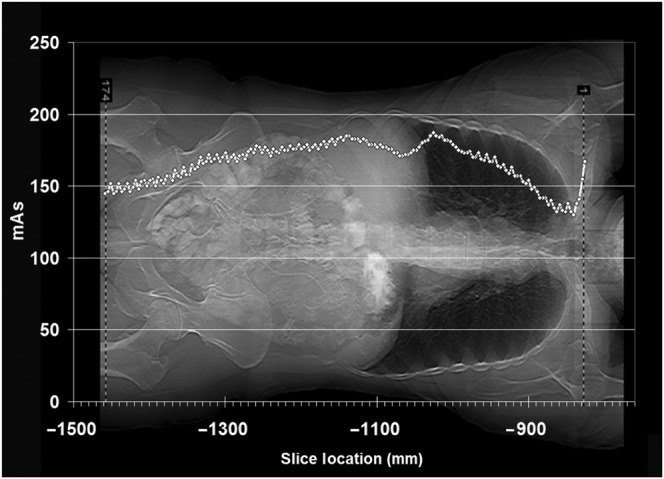
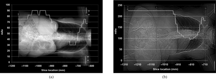
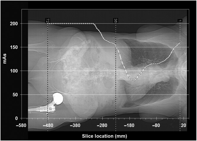
Results: The calculated DLP values were in good agreement (±5%) with the displayed values. The calculated average CDTI(vol) values were in similar agreement with the displayed CTDI(vol) values but only for two of the three scanners. In the other scanner the displayed CTDI(vol) values were found to be overestimated by about 25%. As an additional application of this method the differences among the tube modulation techniques used by the three CT scanners were investigated.
Conclusion: This method is a useful tool for radiation dose surveys.
Figures
Similar articles
-
A comprehensive method for calculating patient effective dose and other dosimetric quantities from CT DICOM images.AJR Am J Roentgenol. 2012 Jul;199(1):133-41. doi: 10.2214/AJR.11.7429. AJR Am J Roentgenol. 2012. PMID: 22733904
-
Dose reduction for abdominal and pelvic MDCT after change to graduated weight-based protocol for selecting quality reference tube current, peak kilovoltage, and slice collimation.AJR Am J Roentgenol. 2013 Jun;200(6):1298-303. doi: 10.2214/AJR.12.9387. AJR Am J Roentgenol. 2013. PMID: 23701068
-
Extraction of tube current values from DICOM CT images for patient dose estimation.Med Phys. 2010 Jun;37(6):2951-5. doi: 10.1118/1.3429054. Med Phys. 2010. PMID: 20632607
-
Utilizing a simple CT dosimetry phantom for the comprehension of the operational characteristics of CT AEC systems.Med Phys. 2013 Nov;40(11):111918. doi: 10.1118/1.4826160. Med Phys. 2013. PMID: 24320451
-
Automated framework for digital radiation dose index reporting from CT dose reports.AJR Am J Roentgenol. 2011 Nov;197(5):1170-4. doi: 10.2214/AJR.11.6650. AJR Am J Roentgenol. 2011. PMID: 22021511
Cited by
-
Open-Source Radiation Exposure Extraction Engine (RE3) with Patient-Specific Outlier Detection.J Digit Imaging. 2016 Aug;29(4):406-19. doi: 10.1007/s10278-015-9852-y. J Digit Imaging. 2016. PMID: 26644157 Free PMC article.
-
Electronic collimation of radiographic images: does it comprise an overexposure risk?Br J Radiol. 2018 Jun;91(1086):20170958. doi: 10.1259/bjr.20170958. Epub 2018 Mar 22. Br J Radiol. 2018. PMID: 29544343 Free PMC article.
-
A comparison of methods to estimate organ doses in CT when utilizing approximations to the tube current modulation function.Med Phys. 2012 Aug;39(8):5212-28. doi: 10.1118/1.4736807. Med Phys. 2012. PMID: 22894446 Free PMC article.
-
Image Quality Comparison between Digital Breast Tomosynthesis Images and 2D Mammographic Images Using the CDMAM Test Object.J Imaging. 2022 Aug 21;8(8):223. doi: 10.3390/jimaging8080223. J Imaging. 2022. PMID: 36005466 Free PMC article.
-
Medical image file formats.J Digit Imaging. 2014 Apr;27(2):200-6. doi: 10.1007/s10278-013-9657-9. J Digit Imaging. 2014. PMID: 24338090 Free PMC article. Review.
References
-
- Lewis MA, Edyvean S. Patient dose reduction in CT. Br J Radiol 2005;78:880–3 - PubMed
-
- Shrimpton PC, Hillier MC, Lewis MA, Dunn M. Doses form computed tomography examinations in the UK—2003 Review. NRPB-W67, National Radiological Protection Board, Chilton, UK, 2005.
-
- ImPACT CT Patient Dosimetry Calculator, Version 1.0. Impact group website. Available from: www.impactscan.org/ctdosimetry.htm. Accessed September 23, 2009.
-
- McCollough CH, Bruesewitz MR, Kofler JM. CT dose reduction and dose management tools: overview of available options. Radiographics 2006;26:503–12 - PubMed
-
- Nicholson R, Fetherston S. Primary radiation outside the imaged volume of a multislice helical CT scan. Br J Radiol 2002;75:518–22 - PubMed
MeSH terms
LinkOut - more resources
Full Text Sources
Medical
Research Materials
