The use of joint-specific and whole-body MRI in osteonecrosis: a study in patients with juvenile systemic lupus erythematosus
- PMID: 21081568
- PMCID: PMC3473496
- DOI: 10.1259/bjr/34972239
The use of joint-specific and whole-body MRI in osteonecrosis: a study in patients with juvenile systemic lupus erythematosus
Abstract
Objective: This study aimed to estimate the prevalence of osteonecrosis (ON) in juvenile systemic lupus erythematosus (SLE) patients using joint-specific and whole-body MRI; to explore risk factors that are associated with the development of ON; and to evaluate prospectively patients 1 year after initial imaging.
Method: Within a 2 year period, we studied 40 juvenile SLE patients (aged 8-18 years) with a history of steroid use of more than 3 months duration. Risk factors including disease activity, corticosteroid use, vasculitis, Raynaud's phenomenon and lipid profile were evaluated. All patients underwent MRI of the hips, knees and ankles using joint-specific MRI. Whole-body STIR (short tau inversion recovery) MRI was performed in all patients with ON lesions.
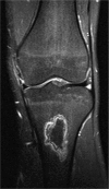
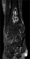
Results: Osteonecrosis was identified in 7 patients (17.5 %) upon joint-specific MRI. Whole-body STIR MRI detected ON in 6 of these 7 patients. There was no significant difference between the ON and non-ON groups in the risk factors studied. One patient had pre-existing symptomatic ON. At 1 year follow-up, the ON lesions had resolved in one patient, remained stable in four and decreased in size in two. No asymptomatic patients with ON developed clinical manifestations.
Conclusion: Whole-body STIR MRI may be useful in detecting ON lesions in juvenile SLE patients but larger studies are needed to define its role.
Figures


References
-
- Mont MA, Hungerford DS. Non-traumatic avascular necrosis of the femoral head. J Bone Joint Surg 1995;77:459–74 - PubMed
-
- Mankin HJ. Nontraumatic necrosis of bone (osteonecrosis). N Engl J Med 1992;326:1473–9 - PubMed
-
- Assouline-Dayan Y, Chang C, Greenspan A, Shoenfeld Y, Gershwin ME. Pathogenesis and natural history of osteonecrosis. Semin Arthritis Rheum 2002;32:92–124 - PubMed
-
- Wang GJ, Sweet DE, Reger SI, Thompson RC. Fat-cell changes as a mechanism of avascular necrosis of the femoral head in cortisone-treated rabbits. J Bone Joint Surg Am 1977;59:729–35 - PubMed
-
- Glueck CJ, Freiberg R, Glueck HI, Henderson C, Welch M, Tracy T, et al. Hypofibrinolysis: a common, major cause of osteonecrosis. Am J Hematol 1994;45:156–66 - PubMed
Publication types
MeSH terms
LinkOut - more resources
Full Text Sources
Medical
Miscellaneous

