. 2011 Mar;79(3):340-50.
doi: 10.1124/mol.110.068478. Epub 2010 Nov 16.

Progesterone receptor membrane component 1 inhibits the activity of drug-metabolizing cytochromes P450 and binds to cytochrome P450 reductase

Affiliations

Progesterone receptor membrane component 1 inhibits the activity of drug-metabolizing cytochromes P450 and binds to cytochrome P450 reductase

Elzbieta Szczesna-Skorupa et al. Mol Pharmacol. 2011 Mar.

Abstract

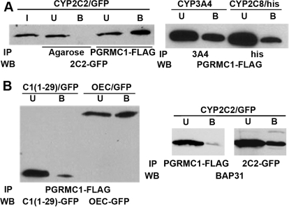
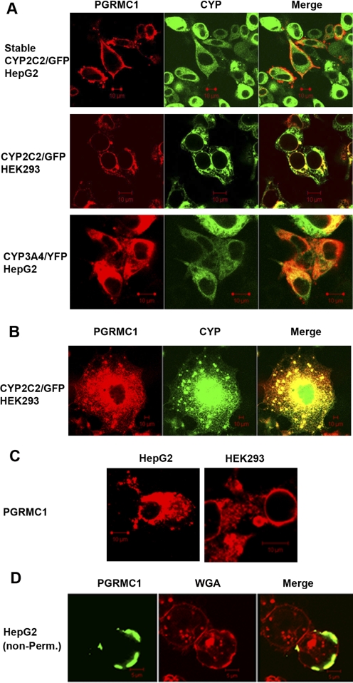
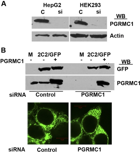
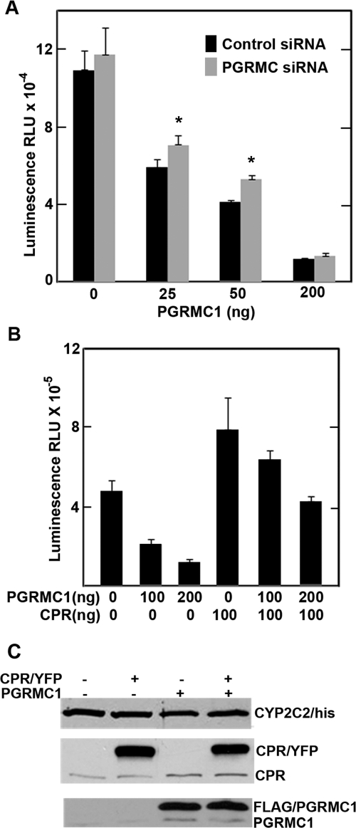
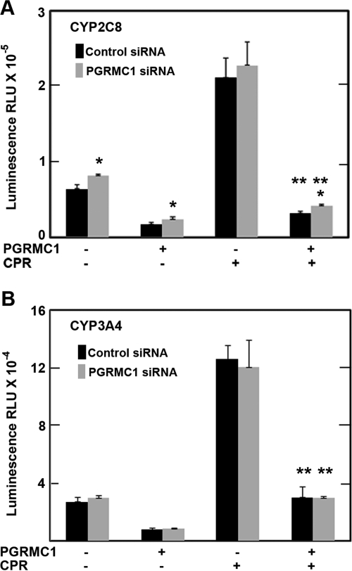
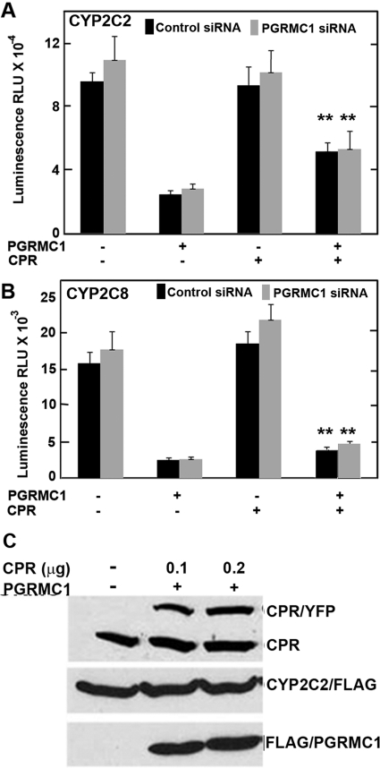
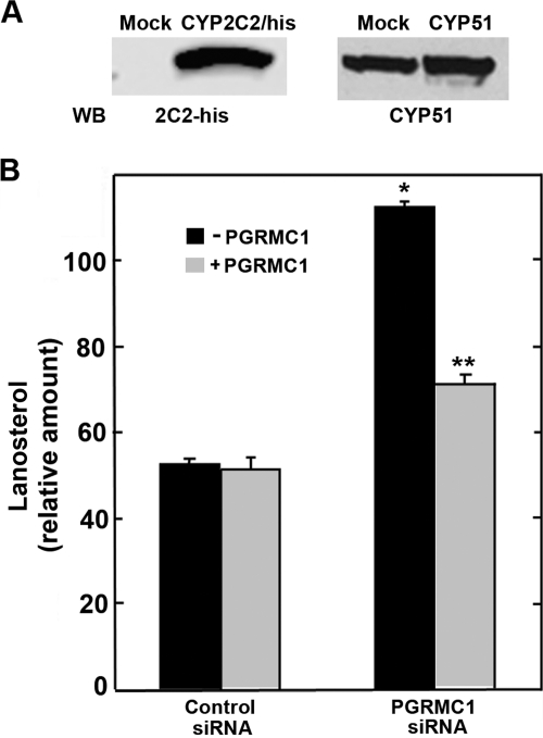
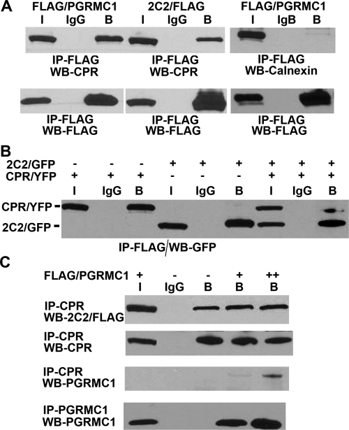
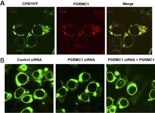
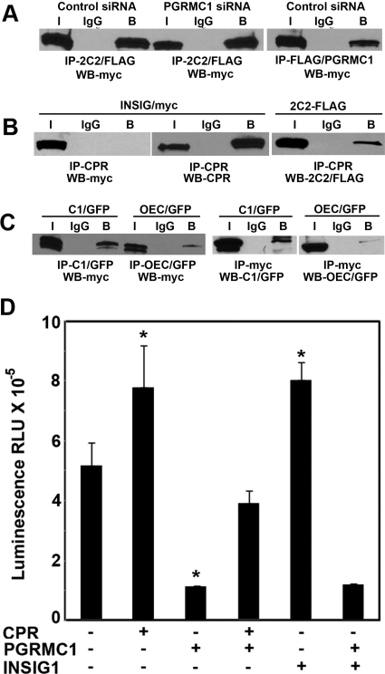
Progesterone receptor membrane component 1 (PGRMC1) has been shown to interact with several cytochromes P450 (P450s) and to activate enzymatic activity of P450s involved in sterol biosynthesis. We analyzed the interactions of PGRMC1 with the drug-metabolizing P450s, CYP2C2, CYP2C8, and CYP3A4, in transfected cells. Based on coimmunoprecipitation assays, PGRMC1 bound efficiently to all three P450s, and binding to the catalytic cytoplasmic domain of CYP2C2 was much more efficient than to a chimera containing only the N-terminal transmembrane domain. Down-regulation of PGRMC1 expression levels in human embryonic kidney 293 and HepG2 cell lines stably expressing PGRMC1-specific small interfering RNA had no effect on the endoplasmic reticulum localization and expression levels of P450s, whereas enzymatic activities of CYP2C2, CYP2C8, and CYP3A4 were slightly higher in PGRMC1-deficient cells. Cotransfection of cells with P450s and PGRMC1 resulted in PGRMC1 concentration-dependent inhibition of the P450 activities, and this inhibition was partially reversed by increased expression of the P450 reductase (CPR). In contrast, CYP51 activity was decreased by down-regulation of PGRMC1 and expression of PGRMC1 in the PGRMC1-deficient cells increased CYP51 activity. In cells cotransfected with CPR and PGRMC1, strong binding of CPR to PGRMC1 was observed; however, in the presence of CYP2C2, interaction of PGRMC1 with CPR was significantly reduced, suggesting that CYP2C2 competes with CPR for binding to PGRMC1. These data show that in contrast to sterol synthesizing P450, PGRMC1 is not required for the activities of several drug-metabolizing P450s, and its overexpression inhibits those P450 activities. Furthermore, PGRMC1 binds to CPR, which may influence P450 activity.


Figures

Fig. 1.
Fig. 1.
Binding of FLAG/PGRMC1 to P450s 2C2, 3A4, and 2C8. A, HEK293 cells were transfected with expression plasmids for FLAG/PGRMC1 and either CYP2C2/GFP, CYP3A4/YFP, or CYP2C8/FLAG/his. After 24 h, cellular lysates were prepared, and FLAG/PGRMC1 was immunoprecipitated with M2-agarose, CYP3A4 with anti-3A4, or CYP2C8/FLAG/his with anti-histidine antisera. Western blots were probed with either anti-GFP or anti-FLAG antibodies. Input (I) and bound (B) and unbound (U) fractions were analyzed. In both A and B, the bound fraction was concentrated 10-fold relative to unbound and input. B, HEK293 cells were transfected with FLAG/PGRMC1 and either C1(1–29)/GFP or OEC/GFP and after 24 h, cellular lysates were prepared and FLAG/PGRMC1 was immunoprecipitated with M2-agarose. Unbound (U) and bound (B) fractions were analyzed by Western blots probed with anti-GFP antibody. C, HEK293 cells were transfected with FLAG/PGRMC1 and CYP2C2/GFP as in A. FLAG/PGRMC1 and CYP2C2/GFP were immunoprecipitated from cellular lysates with M2-agarose and GFP antibodies, respectively, and the amount of BAP31 in the immunoprecipitates was determined by Western blotting with an antibody against BAP31.
Fig. 2.
Fig. 2.
Subcellular localization of PGRMC1 and P450s. A, HepG2 cells stably expressing CYP2C2/GFP were transfected with FLAG/PGRMC1 (1 μg) (top row), HEK293 cells were transiently cotransfected with FLAG/PGRMC1 (0.5 μg) and CYP2C2/GFP (1 μg) (middle row), and HepG2 cells were transiently cotransfected with FLAG/PGRMC1 (1 μg) and CYP3A4/YFP (1 μg) (bottom row). Twenty-four hours after transfection, cells were fixed, permeabilized, and P450s were detected by fluorescence and PGRMC1 by immunostaining with an anti-FLAG antibody followed by Cy5-conjugated secondary antibody. B, a higher magnification image of HEK293 cell transiently cotransfected with CYP2C2/GFP and FLAG/PGRMC1 and processed as in A. C, HepG2 or HEK293 cells were transiently transfected with FLAG/PGRMC1 only and processed as in A. D, HepG2 cells were transfected with FLAG/PGRMC1 (1 μg), and after 24 h, cells were fixed and immunostained with anti-FLAG antibody or with wheat germ agglutinin (a plasma membrane marker) without permeabilization. Cells were analyzed by confocal microscopy. Scale bars, 10 μm.
Fig. 3.
Fig. 3.
Down-regulation of PGRMC1 expression in HEK293 and HepG2 cells. A, lysates from HEK293 and HepG2 cells stably expressing either nonsilencing control siRNA (C) or PGRMC1 specific siRNA (si) were prepared, and PGRMC1 or actin was detected by Western analysis with an antibodies against PGRMC1 or actin, respectively. B, HEK293 cells stably expressing either control or PGRMC1 specific siRNA were transfected with expression plasmids for CYP2C2/GFP with or without FLAG/PGRMC1, and after 24 h, cellular lysates were prepared, and CYP2C2/GFP and PGRMC1 were detected by Western analysis using antisera to GFP and PGRMC1, respectively (top). Both endogenous (bottom band) and transfected FLAG-tagged PGRMC1 (top band) were detected. M indicates mock-transfected cells. The lower panel shows HEK293 cells stably expressing either control or PGRMC1 siRNA transfected with CYP2C2/GFP fixed and imaged by fluorescent microscopy.
Fig. 4.
Fig. 4.
PGRMC1 effect on the activity of CYP2C2 in HEK293 cells. A, HEK293 cells stably expressing either control (■) or PGRMC1 siRNA (▩) were plated into 12-well plates, and 24 h later, cells were transfected in triplicate either with an expression vector for CYP2C2/FLAG (0.5 μg/well) alone or additionally with indicated amounts of FLAG/PGRMC1 expression vector. After 24 h, the enzymatic activity of CYP2C2 was measured using the P450-Glo assay as described under Materials and Methods. B, HEK293 cells were transfected with an expression vector for CYP2C2/FLAG (0.5 μg/well) alone or with FLAG/PGRMC1 and 0.1 μg of CPR/YFP expression vectors. The activity of CYP2C2 was assayed 24 h after transfection. Background luminescence from mock-transfected cells was subtracted from all samples. Data were analyzed by the Student's t test. *, significant difference between the PGRMC siRNA sample and the control siRNA sample, p < 0.05, n = 3. A and B, the mean value from triplicates and the standard deviation are shown. C, HEK293 cells were transfected with CYP2C2/FLAG (0.5 μg), CPR/YFP (0.1 μg), and FLAG/PGRMC1 (0.2 μg), as indicated, and after 24 h, cells were lysed in radioimmunoprecipitation assay buffer, and respective proteins were detected by Western analysis. Both endogenous (bottom bands) and transfected CPR/YFP or FLAG/PGRMC1 (top bands) were detected as indicated.
Fig. 5.
Fig. 5.
PGRMC1 effect on the activities of CYP2C8 and CYP3A4 in HEK293 cells. HEK293 cells stably expressing control (■) or PGRMC1 siRNA (▩) were transfected as in Fig. 4 either with 0.5 μg per well of CYP2C8/FLAG/His (A) or CYP3A4 (B) expression vectors with or without 0.2 μg of FLAG/PGRMC1 and 0.1 μg of CPR/YFP. Enzymatic activity was assayed after 24 h as described in the legend to Fig. 4. Data were analyzed by the Student's t test. *, significant difference between the PGRMC1 siRNA sample and the control siRNA sample; and **, significant difference between cells expressing CPR and PGRMC1 compared with cells expressing only PGRMC1, p < 0.05, n = 3.
Fig. 6.
Fig. 6.
PGRMC1 effect on CYP2C2 and CYP2C8 activity and expression levels in transfected HepG2 cells. A and B, HepG2 cells stably expressing control (■) or PGRMC1 siRNA (▩) were transfected as in Fig. 4 with 1 μg per well of CYP2C2/FLAG (A) or 1 μg of CYP2C8/FLAG/his (B) (compared with 0.2 μg in Fig. 4) expression vectors with or without 0.1 μg of CPR/YFP and either 1 μg (A) or 0.5 μg (B) of FLAG/PGRMC1. Activities were assayed after 24 h as described in the legend to Fig. 4. C, transfected HepG2 cells were lysed in radioimmunoprecipitation assay buffer, and CPR (top), CYP2C2 (middle), and PGRMC1 (bottom) were detected by Western analysis. Both endogenous (bottom band) and transfected CPR/YFP (top band) were detected. **, significant difference between cells expressing CPR and PGRMC1 compared with cells expressing only PGRMC1, p < 0.05, n = 3.
Fig. 7.
Fig. 7.
PGRMC1 effect on lanosterol levels in HEK293 cells. A, HEK293 cells were transfected with CYP2C2/his (left) or CYP51 (right), and 24 h later cellular lysates were analyzed by Western analysis with an anti-histidine or anti-CYP51 antibodies. B, HEK293 cells stably expressing control or PGRMC1 siRNA were either mock-transfected (■) or transfected with a FLAG/PGRMC1 expression plasmid (▩) and 24 h later were incubated in DMEM containing 5% lipoprotein-deficient serum and 40 mM mevalonate for 10 h. Extracted sterols were analyzed with GC/MS as described under Materials and Methods. The mean values and standard deviations from triplicates are shown. *, significant difference between PGRMC1 siRNA and control siRNA samples; **, significant difference between cells expressing PGRMC1 with cells not expressing PGRMC1, p < 0.05, n = 3.
Fig. 8.
Fig. 8.
Binding of PGRMC1 to CPR. A, HEK293 cells were transfected with expression vectors for CPR/YFP and either FLAG/PGRMC1 or CYP2C2/FLAG (left) or FLAG/PGRMC1 and calnexin (right) and after 24 h, cellular lysates were subjected to immunoprecipitation with M2-agarose or agarose-conjugated nonimmune IgG. CPR in the immunoprecipitates was detected by Western analysis using an antibody against CPR and calnexin with an anti-calnexin antibody (top). Bottom, controls are shown in which FLAG immunoprecipitates were analyzed by Western blot with FLAG antibody. Bound (B) fractions are concentrated 5-fold compared with input (I) fractions. B, HEK293 cells were transfected with expression vectors for FLAG/PGRMC1 and either CYP2C2/GFP, CPR/YFP, or both, as indicated. After 24 h, lysates from transfected cells were analyzed by immunoprecipitation with M2-agarose or agarose-conjugated IgG and CYP2C2/GFP and CPR/YFP were detected in the immunoprecipitates by Western analysis with an antibody against GFP, which recognizes both GFP and YFP. C, HEK293 cells were cotransfected with expression vectors for CYP2C2/FLAG/his and CPR/YFP without or with 0.5 μg (+) or 1 μg (++) of FLAG/PGRMC1. After 24 h, CPR was immunoprecipitated from cellular lysates with an antibody against CPR, and PGRMC1 was immunoprecipitated with an antibody against PGRMC1. CYP2C2/FLAG/His (top), CPR/YFP (top middle), and FLAG/PGRMC1 (bottom middle and bottom) in the immunoprecipitates were detected by Western analysis with an anti-FLAG, anti-CPR, and anti-PGRMC1 antibodies, respectively. A to C, at least three experiments were performed, and representative data for each are shown.
Fig. 9.
Fig. 9.
Subcellular localization of CPR in cells expressing control and PGRMC1 siRNA. A, HepG2 cells were transfected with expression vectors for CPR/YFP and FLAG/PGRMC1, and after 24 h, fixed cells were permeabilized and subjected to immunostaining with anti-FLAG antibody followed by Cy5-conjugated secondary antibody to detect PGRMC1 (red). B, control or PGRMC1-deficient HEK293 cells were transfected with expression vectors for CPR/YFP only or with CPR/YFP and FLAG/PGRMC1 (far right). Fixed cells were analyzed by confocal microscopy. Scale bars, 10 μm.
Fig. 10.
Fig. 10.
INSIG1 binds CYP2C2 and stimulates its activity. A, HEK293 cells expressing control or PGRMC1 siRNA were transfected with expression vectors for INSIG1/myc and either CYP2C2/FLAG/his (left) or FLAG/PGRMC1 (right). Twenty-four hours later, cellular lysates were immunoprecipitated with M2-agarose or agarose-conjugated IgG, and INSIG1/myc in the immunoprecipitates was detected by Western analysis with anti-myc antibody. B, lysates of HEK293 cells cotransfected with CPR/YFP and either INSIG/myc or CYP2C2/FLAG, as indicated, were immunoprecipitated with an antibody against CPR, and Western blots were probed with anti-myc antibody to detect INSIG/myc (left), anti-CPR to detect CPR (middle), or anti-FLAG to detect CYP2C2/FLAG. C, lysates of HEK293 cells cotransfected with INSIG/myc and either C1(1–29)/GFP or OEC/GFP, as indicated, were immunoprecipitated with an antibody against GFP or agarose-conjugated IgG and INSIG/myc was detected on Western blots with an anti-myc antibody (left). Right, identical lysates immunoprecipitated with an anti-myc antibody and probed with an antibody against GFP. For A to C, bound (B) fractions were concentrated 5-fold compared with input (I) fractions. Similar results were obtained in three independent experiments. D, CYP2C2 activity in the presence of INSIG1. HEK293 cells grown in 12-well plates were transfected with 0.5 μg of CYP2C2/FLAG/His with or without addition of CPR/YFP (0.1 μg), FLAG/PGRMC1 (0.2 μg), and INSIG1/myc (0.2 μg per well). Activities were determined after 24 h as described in the legend to Fig. 4. Background luminescence from mock-transfected cells was subtracted from all samples. The mean value from triplicates and the standard deviation are shown. *, significant differences between cells expressing the indicated protein and untransfected cells, p < 0.05, n = 3.

Similar articles

Cited by

References

    1. Ahmed IS, Rohe HJ, Twist KE, Mattingly MN, Craven RJ. (2010) Progesterone receptor membrane component 1 (Pgrmc1): a heme-1 domain protein that promotes tumorigenesis and is inhibited by a small molecule. J Pharmacol Exp Ther 333:564–573 - PubMed
    1. Ahn K, Szczesna-Skorupa E, Kemper B. (1993) The amino-terminal 29 amino acids of cytochrome P450 2C1 are sufficient for retention in the endoplasmic reticulum. J Biol Chem 268:18726–18733 - PubMed
    1. Backes WL, Kelley RW. (2003) Organization of multiple cytochrome P450s with NADPH-cytochrome P450 reductase in membranes. Pharmacol Ther 98:221–233 - PubMed
    1. Bridges A, Gruenke L, Chang YT, Vakser IA, Loew G, Waskell L. (1998) Identification of the binding site on cytochrome P450 2B4 for cytochrome b5 and cytochrome P450 reductase. J Biol Chem 273:17036–17049 - PubMed
    1. Cahill MA. (2007) Progesterone receptor membrane component 1: an integrative review. J Steroid Biochem Mol Biol 105:16–36 - PubMed

Publication types

MeSH terms