. 2011 Jan;62(3):1255-69.
doi: 10.1093/jxb/erq354. Epub 2010 Nov 16.

Nuclear activity of sperm cells during Hyacinthus orientalis L. in vitro pollen tube growth

Affiliations

Nuclear activity of sperm cells during Hyacinthus orientalis L. in vitro pollen tube growth

Krzysztof Zienkiewicz et al. J Exp Bot. 2011 Jan.

Abstract

In this study, the transcriptional state and distribution of RNA polymerase II, pre-mRNA splicing machinery elements, and rRNA transcripts were investigated in the sperm cells of Hyacinthus orientalis L. during in vitro pollen tube growth. During the second pollen mitosis, no nascent transcripts were observed in the area of the dividing generative cell, whereas the splicing factors were present and their pools were divided between newly formed sperm cells. Just after their origin, the sperm cells were shown to synthesize new RNA, although at a markedly lower level than the vegetative nucleus. The occurrence of RNA synthesis was accompanied by the presence of RNA polymerase II and a rich pool of splicing machinery elements. Differences in the spatial pattern of pre-mRNA splicing factors localization reflect different levels of RNA synthesis in the vegetative nucleus and sperm nuclei. In the vegetative nucleus, they were localized homogenously, whereas in the sperm nuclei a mainly speckled pattern of small nuclear RNA with a trimethylguanosine cap (TMG snRNA) and SC35 protein distribution was observed. As pollen tube growth proceeded, inhibition of RNA synthesis in the sperm nuclei was observed, which was accompanied by a gradual elimination of the splicing factors. In addition, analysis of rRNA localization indicated that the sperm nuclei are likely to synthesize some pool of rRNA at the later steps of pollen tube. It is proposed that the described changes in the nuclear activity of H. orientalis sperm cells reflect their maturation process during pollen tube growth, and that mature sperm cells do not carry into the zygote the nascent transcripts or the splicing machinery elements.


Figures
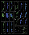
Fig. 1.
Fig. 1.
Localization of the incorporated 5-bromouracil (green colour) in hyacinth pollen tubes. (A1–A3) A pollen tube after 8 h of growth containing the dividing generative cell. A strong signal is present in the vegetative nucleus and in the pollen tube cytoplasm. The area of the dividing generative nucleus is completely devoid of fluorescence. (B1–B3) A pollen tube soon after sperm cell formation. The vegetative nucleus exhibits a significantly stronger signal in comparison with the sperm cells. In the sperm nuclei, a few spots of the signal are present. (C1–C3) After 12 h of pollen tube growth, the sperm nuclei exhibit a signal intensity comparable with that observed in the vegetative nucleus. (D1–D3) In both sperm nuclei of the pollen tube growing for 24 h, the signal corresponding to nascent transcripts is localized in the form of numerous irregular fluorescent clusters. (E1–E3) A pollen tube growing for 32 h. Strong homogenous fluorescence is visible in the vegetative nucleus as well as in the pollen tube cytoplasm. In the area of the sperm nuclei, only a few spots of weak signal are present. (F1–F3) No labelling can be observed in the sperm nuclei after 36 h of pollen tube growth. Weak fluorescence is present in the pollen tube cytoplasm. (G1 and G2) Control reaction performed on pollen tubes treated with actinomycin D. (H1 and H2) Positive control showing the presence of nascent transcripts in the cells of the young anther wall. An intense signal is present in the nuclei and cytoplasm of the parenchyma cells, whereas the vacuoles are devoid of green fluorescence; values presented in the upper right corner of the figures indicate the percentage of pollen tubes exhibiting the labelling pattern shown on the image. Z-series images corresponding to the whole volume of growing pollen tubes from three independent experiments (n=50) were captured and used for statistical analysis. VN, vegetative nucleus; GN, generative nucleus; SN, sperm nucleus; N, nucleus; V, vacuole; bars=10 μm.
Fig. 2.
Fig. 2.
Profile of total RNA in the growing pollen tubes of hyacinth. (A) Ethidium bromide-stained gel of total RNA isolated from pollen at maturity (MP), after hydration (H), and after 1, 3, 6, 12, 24, and 36 h of in vitro germination. Well-distinguishable bands corresponding to 26S rRNA and 18S rRNA as well as to tRNA are indicated with arrows. (B) Densitometric data corresponding to the 26S rRNA (black bars), 18S rRNA (grey bars), and tRNA (white bars) from A. The values represent the average of thee replicate experiments; the error bars indicate 1 SEM.
Fig. 3.
Fig. 3.
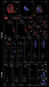
Localization of rRNA (red colour) in hyacinth pollen tubes by FISH. (A1–A3) In the young pollen tube, the signal corresponding to 26S rRNA is observed only in the cytoplasm. Accumulation of fluorescence occurs inside the pollen grain and at the tip of the small pollen tube (arrow). Pollen nuclei do not show any labelling. (B1 and B2) After 3 h of pollen tube growth, the strongest signal is present in the pollen tube cytoplasm. At this time, the fluorescence corresponding to 26S rRNA appears in the vegetative nucleus, whereas the generative nucleus does not show any fluorescence. (B3–B6) On the consecutive optical sections of both pollen tube nuclei (area marked with the dashed line on B1 and B2), accumulation of the signal in the vegetative nucleus is observed in a round-shaped fluorescent cluster (arrowhead). (C1–C3) After sperm cell formation, 26S rRNA appears to be located in both sperm nuclei and the vegetative nucleus. The arrowhead indicates the fragment of the other pollen tube. (C4–C6) Magnification of the pollen tube zone containing sperm cells (area marked with the dashed line on C1). In both sperm nuclei, the signal is present in the form of irregular fluorescent clusters, which appear to occupy nuclear areas devoid of DNA (C6; circles). (D1–D4) The fluorescence corresponding to immature rRNAs is observed only in the sperm nuclei where it is visible as numerous spots of different sizes and shapes (D4). (E1-E3) A positive control performed on the somatic tissues of hyacinth anther revealed accumulation of ITS containing rRNAs exclusively in the nucleoli of the parenchyma cell nuclei (arrowheads); values presented in the upper right corner of the figures indicate the percentage of pollen tubes exhibiting the labelling pattern showed on the image. Z-series images corresponding to the whole volume of growing pollen tubes from three independent experiments (n=50) were captured and used for statistical analysis. VN, vegetative nucleus; GN, generative nucleus; SN, sperm nucleus; N, nucleus; bars=10 μm.
Fig. 4.
Fig. 4.
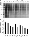
Analysis of protein contents during H. orientalis pollen tube growth. (A) Coomassie blue-stained gel of total proteins from pollen grains at maturity (MP), after hydration (H), and after 1, 3, 6, 12, 24, and 36 h of in vitro germination. Lanes marked as 6hCHX and 12hCHX indicate pollen tubes treated for 3 h with cycloheximide added to the medium at the third and ninth hour of growth, respectively. Lanes 6hC and 12hC indicate control pollen tubes growing for 6 h and 12 h, respectively. (B) Protein concentration (μg) in 1 μl of extracts isolated from the mature, hydrated, and germinating pollen grains of hyacinth. The temporal variants of pollen tube growth are the same as shown in A. Grey blocks of the graph correspond to the pollen tubes treated with cycloheximide. The values represent the average of three replicate experiments; the error bars indicate 1 SEM.
Fig. 5.
Fig. 5.
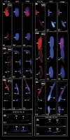
Localization of hypo- (Pol IIA) and hyperphosphorylated (Pol IIO) forms of RNA polymerase II (red colour) in hyacinth pollen tubes. (A1–A3) In the pollen tube growing for 8 h, the labelling indicating Pol IIA is present exclusively in the area of the vegetative and generative nuclei. In the latter nucleus, only some irregular fluorescence clusters are present. In the vegetative nucleus, the labelling is much more intense and uniformly distributed throughout the nucleoplasm. (B1–B3) After generative cell division, both the sperm nuclei exhibit the presence of irregular fluorescent spots corresponding to Pol IIA. The magnification of the area marked with the dashed line in B1 is shown in Supplementary Fig. S5 at JXB online. (C1-C3) In the pollen tube growing for 36 h, both sperm cells show no labelling. A strong signal is present in the vegetative nucleus. (D1 and D2). Control reaction by omitting the primary anti-Pol IIA antibody. (E1–E3) Before generative cell division, the strong and uniformly distributed fluorescence indicating Pol IIO is located in the vegetative nucleus. In the generative nucleus, numerous clusters of less intense signal are present. (F1–F3) After second pollen mitosis, the signal is observed in the vegetative and sperm nuclei, wherein numerous spots of fluorescence are present. (G1–G3) Later steps of pollen tube growth were accompanied by a lack of the signal in the sperm nuclei and its reduction in the vegetative nucleus. (H1 and H2) Control reaction conducted by omitting incubation with the anti-Pol IIO antibody; values presented in the upper right corner of the figures indicate the percentage of pollen tubes exhibiting the labelling pattern showed on the image. Z-series images corresponding to the whole volume of growing pollen tubes from three independent experiments (n=50) were captured and used for statistical analysis; VN, vegetative nucleus; GN, generative nucleus; SN, sperm nucleus; bars=10 μm.
Fig. 6.
Fig. 6.
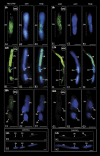
Immunolocalization of pre-mRNA splicing machinery elements (TMG snRNA and SC35 protein; green colour) in hyacinth pollen tubes. (A1–A3) A dividing generative cell. The signal appears to be located between two groups of daughter chromosomes. (B1–B3) After 12 h of pollen tube growth, the fluorescence corresponding to TMG snRNAs is present in two sperm nuclei. The signal is visible as numerous irregular fluorescent spots spread throughout the sperm nucleoplasm. In the vegetative nucleus, strong and uniform fluorescence is observed. Magnification of the area marked with a dashed line on B1 is shown in Supplementary Fig. S6 at JXB online. (C1–C3) A pollen tube growing for 36 h. A weak signal is observed only in the vegetative nucleus, whereas both sperm nuclei are almost completely devoid of labelling. (D1 and D2) Control reaction by omitting the primary anti-TMG antibody. (E1–E3) A generative cell during mitosis. The signal indicating the SC35 splicing factor is located in the area between the newly forming sperm nuclei. (F1–F3) After 12 h of pollen tube growth, the signal is present in the vegetative nucleus as well as in both sperm nuclei. In the vegetative nucleus, intense and uniformly distributed fluorescence is observed. Sperm nuclei exhibit low intensity staining, localized mainly in the form of numerous irregular fluorescent clusters. Magnification of the area marked with the dashed line on F1 is shown in Supplementary Fig. S7. (G1–G3) A pollen tube growing for 36 h. Lack of the signal corresponding to SC35 protein is observed in the sperm nuclei and in the vegetative nucleus. (H1 and H2) Control reaction by omitting incubation with the primary anti-SC35 antibody. The values presented in the upper right corner of the figures indicate the percentage of pollen tubes exhibiting the labelling pattern shown in the image. Z-series images corresponding to the whole volume of growing pollen tubes from three independent experiments (n=50) were captured and used for statistical analysis; VN, vegetative nucleus; GN, generative nucleus; SN, sperm nucleus; bars=10 μm.

Similar articles

Cited by

References

    1. Bayer M, Nawy T, Giglione C, Galli M, Meinnel T, Lukowitz W. Paternal control of embryonic patterning in Arabidopsis thaliana. Science. 2009;323:1485–1488. - PubMed
    1. Becker JD, Boavida LC, Carneiro J, Haury M, Feijó JA. Transcriptional profiling of Arabidopsis tissues reveals the unique characteristic of the pollen transcriptome. Plant Physiology. 2003;133:713–725. - PMC - PubMed
    1. Bentley DL. Rules of engagement: co-transcriptional recruitment of pre-mRNA processing factors. Current Opinion in Cell Biology. 2005;17:251–256. - PubMed
    1. Berger F, Hamamura Y, Ingouff M, Higashiyama T. Double fertilization—caught in the act. Trends in Plant Science. 2008;13:437–443. - PubMed
    1. Bino RJ, Van Tuyl JM, De Vries JN. Flow cytometric determination of relative nuclear DNA contents in bicellulate and tricellulate pollen. Annals of Botany. 1990;65:3–8.

Publication types