. 2010 Dec 14;107(50):21248-55.
doi: 10.1073/pnas.1015855107. Epub 2010 Nov 16.

Granulocyte-colony stimulating factor promotes lung metastasis through mobilization of Ly6G+Ly6C+ granulocytes

Affiliations

Granulocyte-colony stimulating factor promotes lung metastasis through mobilization of Ly6G+Ly6C+ granulocytes

Marcin Kowanetz et al. Proc Natl Acad Sci U S A. .

Abstract

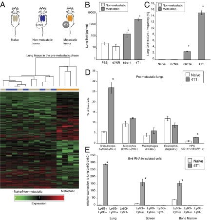
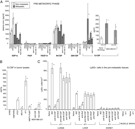
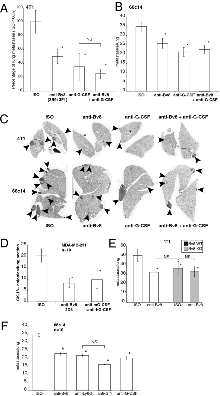
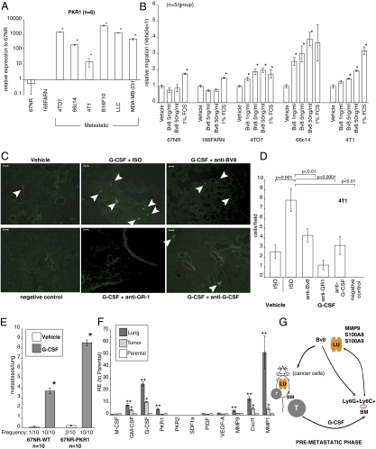
Priming of the organ-specific premetastatic sites is thought to be an important yet incompletely understood step during metastasis. In this study, we show that the metastatic tumors we examined overexpress granulocyte-colony stimulating factor (G-CSF), which expands and mobilizes Ly6G+Ly6C+ granulocytes and facilitates their subsequent homing at distant organs even before the arrival of tumor cells. Moreover, G-CSF-mobilized Ly6G+Ly6C+ cells produce the Bv8 protein, which has been implicated in angiogenesis and mobilization of myeloid cells. Anti-G-CSF or anti-Bv8 antibodies significantly reduced lung metastasis. Transplantation of Bv8 null fetal liver cells into lethally irradiated hosts also reduced metastasis. We identified an unexpected role for Bv8: the ability to stimulate tumor cell migration through activation of one of the Bv8 receptors, prokineticin receptor (PKR)-1. Finally, we show that administration of recombinant G-CSF is sufficient to increase the numbers of Ly6G+Ly6C+ cells in organ-specific metastatic sites and results in enhanced metastatic ability of several tumors.


Conflict of interest statement

The authors declare no conflict of interest.

Figures

Fig. 1.
Fig. 1.
Bv8 is strongly up-regulated in premetastatic lungs of mice bearing metastatic tumors. (A) Design and results of the microarray study comparing gene expression in lungs from BALB/c naïve mice and mice bearing nonmetastatic tumors 67NR and metastatic 4T1 tumors. Lungs from different experimental groups are color-coded (gray, from naïve mice; blue, from 67NR-bearing mice; orange, from 4T1-bearing mice). Note that the metastatic gene expression profile is clearly separated from both naïve and nonmetastatic profiles. Each profile column represents one individual mouse. (B) Bv8 protein concentrations in the premetastatic lungs of mice bearing various tumors (n = 3 per group). (C) FACS analysis of Cd11b+Gr1+ cells in the premetastatic lungs of BALB/c mice bearing various tumors. Asterisk indicates significant difference relative to the nonmetastatic group. (D) FACS analysis of different cell populations in premetastatic lungs from mice bearing 4T1 tumors 2 wk after tumor inoculation. (E) Bv8 expression in Ly6GLy6C cell subpopulations isolated from lungs of naïve or 4T1 tumor-bearing mice. In D and E, asterisk indicates significant difference relative to naïve group. Graphs present means ± SEM.
Fig. 2.
Fig. 2.
Increased G-CSF and Bv8 levels are associated with a metastatic phenotype. (A) Plasma levels of various cytokines in the premetastatic phase in mice bearing nonmetastatic or metastatic tumors. In samples from mice with MDA-MB-231 tumors, m indicates mouse, and h is human. (B) Levels of G-CSF in whole-tumor extracts isolated from mice in the premetastatic phase. (C) Numbers of Ly6G+ cells (per millimeter squared) in various organs during the premetastatic phase in mice bearing 4T1 tumors. Asterisk indicates significant difference relative to naïve group; two asterisks indicate significant difference relative to isotype control (ISO) group (C). Data shown are means ± SEM.
Fig. 3.
Fig. 3.
Neutralization of G-CSF or Bv8 inhibits metastasis. (A) Number of metastases in lungs of mice bearing 4T1 tumors and treated with indicated antibodies (n = 10) for 5.5 wk after tumor inoculation. (B) Number of metastases per lung in mice bearing 66c14 tumors 6 wk after tumor inoculation. Tumors were implanted, and treatment was performed as in A (n = 10). (C) Representative images of lung sections from mice bearing 4T1 or 66c14 tumors and treated with the indicated antibodies. Sections correspond to A (4T1 tumors) or B (66c14 tumors). Arrowheads indicate metastases. (D) Average numbers of CK-18–positive tumor colonies per lung section of SCID/bg mice bearing MDA-MB-231-X1.1 tumors and treated with indicated antibodies for 7 wk. (E) Number of lung metastases in mice transplanted with either Bv8 WT (white bars) or Bv8 KO (gray bars) fetal liver cells and treated with indicated antibodies. (F) Numbers of lung metastases in mice bearing 66c14 tumors and treated with indicated antibody. Asterisk indicates significant difference relative to ISO group. Data shown are means ± SEM.
Fig. 4.
Fig. 4.
G-CSF promotes premetastatic priming and enhances the metastatic potential of several tumors. (A) FACS analysis of Cd11b+Gr1+ cells in tissues isolated from mice pretreated with vehicle or rG-CSF. (B) Representative images of lung sections collected from mice pretreated with vehicle or G-CSF as in A and stained with anti-Ly6G antibody. (Scale bar: 50 μm.) (C) Bv8 levels measured by ELISA in tissues matching those in A. (D) Number of metastases in lungs of mice pretreated with vehicle or rG-CSF and injected i.v. with indicated tumor cells. Representative H&E lung sections (66c14 and 4T1) or images of whole lungs (B16F10) from each group are shown below the graphs. (E) Number of tumors in lungs of BALB/c mice i.v. injected with nonmetastatic 67NR cells and pretreated with human rG-CSF as in C. Frequency denotes numbers of mice with detectable tumors in lungs. (F) Numbers of metastases in lungs of mice injected i.v. with 66c14 cells and treated daily with vehicle or mouse rG-CSF and indicated antibody. Asterisk indicates significant difference compared with Vehicle (AE) or Vehicle/ISO (F) groups. Two asterisks indicate significant difference compared with G-CSF/ISO group. Data shown are means ± SEM.
Fig. 5.
Fig. 5.
Bv8 mediates G-CSF induced metastasis through enhancement of tumor cell migration. (A) qRT-PCR analysis of PKR1 expression by cancer cells in vitro. (B) In vitro transwell migration of cancer cells in response to Bv8. (C) In vivo extravasation of 4T1 cells in BALB/c nude mice pretreated with rG-CSF and indicated antibodies. White arrowheads indicate 4T1 tumor cells in lungs. (Scale bar: 50 μm.) (D) Quantification of the in vivo extravasation assay from C. (E) Number of tumors in lungs of BALB/c mice pretreated with rG-CSF and i.v. injected with 67NR or 67NR-PKR1 cells. Frequency denotes numbers of mice with detectable tumors in lungs. In AE, asterisk indicates significant difference compared with 67NR cells (A), untreated group (B), or vehicle (E). (F) Gene expression analysis of MDA-MB-231 cells isolated either from lung metastases (Lung) or primary tumors (Tumor). Asterisk indicates significant difference compared with parental cell line; double asterisks indicate significant difference compared with Tumor or parental cell lines. (G) Schematic model of the role of G-CSF and Bv8 in metastasis. LU, lung; T, primary tumor; BM, bone marrow. Data are means ± SEM.

Similar articles

Cited by

References

    1. Nguyen DX, Bos PD, Massague J. Metastasis: From dissemination to organ-specific colonization. Nat Rev Cancer. 2009;9:274–284. - PubMed
    1. Psaila B, Lyden D. The metastatic niche: Adapting the foreign soil. Nat Rev Cancer. 2009;9:285–293. - PMC - PubMed
    1. Kaplan RN, et al. VEGFR1-positive haematopoietic bone marrow progenitors initiate the pre-metastatic niche. Nature. 2005;438:820–827. - PMC - PubMed
    1. Hiratsuka S, Watanabe A, Aburatani H, Maru Y. Tumour-mediated upregulation of chemoattractants and recruitment of myeloid cells predetermines lung metastasis. Nat Cell Biol. 2006;8:1369–1375. - PubMed
    1. Kim S, et al. Carcinoma-produced factors activate myeloid cells through TLR2 to stimulate metastasis. Nature. 2009;457:102–106. - PMC - PubMed

MeSH terms