. 2011 Mar;157(Pt 3):793-804.
doi: 10.1099/mic.0.041996-0. Epub 2010 Nov 16.

Induction of cell death after localization to the host cell mitochondria by the Mycobacterium tuberculosis PE_PGRS33 protein

Affiliations

Induction of cell death after localization to the host cell mitochondria by the Mycobacterium tuberculosis PE_PGRS33 protein

Nathalie Cadieux et al. Microbiology (Reading). 2011 Mar.

Abstract

PE_PGRS33 is the most studied member of the unique PE family of mycobacterial proteins. These proteins are composed of a PE domain (Pro-Glu motif), a linker region and a PGRS domain (polymorphic GC-rich-repetitive sequence). Previous studies have shown that PE_PGRS33 is surface-exposed, constitutively expressed during growth and infection, involved in creating antigenic diversity, and able to induce death in transfected or infected eukaryotic cells. In this study, we showed that PE_PGRS33 co-localizes to the mitochondria of transfected cells, a phenomenon dependent on the linker region and the PGRS domain, but not the PE domain. Using different genetic fusions and chimeras, we also demonstrated a direct correlation between localization to the host mitochondria and the induction of cell death. Finally, although all constructs localizing to the mitochondria did induce apoptosis, only the wild-type PE_PGRS33 with its own PE domain also induced primary necrosis, indicating a potentially important role for the PE domain. Considering the importance of primary necrosis in Mycobacterium tuberculosis dissemination during natural infection, the PE_PGRS33 protein may play a crucial role in the pathogenesis of tuberculosis.


Figures
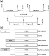
Fig. 1.
Fig. 1.
Protein fusions constructed in this study. (a) Schematic of the method used to construct the fusions. Inserts were engineered to have a HindIII at their 5′ end and a BamHI site at their 3′ end, for cloning into pJW4303. Additional sites such as NruI or EcoRV were included internally, as well as a removable stop codon to allow inserts to be fused together, in-frame. (b) Schematic of the fusions constructed using this method.
Fig. 2.
Fig. 2.
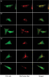
PE_PGRS33 co-localizes to the mitochondria of transfected host cells. RD cells were transfected with the pJW4303 vector (a) or PE_PGRS33 (b and c). Cells stained with the PE_PGRS33-specific mAb 7C4.1F7 (left) and the mitochondria-specific dye MitoTracker Red (centre) and the merged images (right) are shown. Magnification, ×1000; bar, 10 μm.
Fig. 3.
Fig. 3.
Immunofluorescence showing the localization of the different protein fusions in transfected cells. RD cells were transfected with PE_PGRS33 (a), PGRS (b), MaPE–PGRS (c), PE(Δlinker)PGRS (d), MaPE(linker)PGRS (e) and linker–PGRS (f). Cells stained with the PE_PGRS33-specific mAb 7C4.1F7 (left) and the mitochondria-specific dye MitoTracker Red (centre) and the merged images (right) are shown. Magnification, ×1000; bar, 10 μm.
Fig. 4.
Fig. 4.
Time-course showing the percentage of live, apoptotic and necrotic cells as determined by a combination of Hoechst 33342, YO-PRO-1 and PI staining, using the Vybrant Apoptosis Assay kit. T-REx RD cells were collected, stained and analysed by flow cytometry at different times following induction of protein expression. Based on differential staining with Hoechst and PI, the cells were categorized into the following subpopulations: vital (V) cells that stained brightly with Hoechst, but were negative for PI, indicating healthy plasma membranes; apoptotic (Ap) cells at mid (M-Ap), late (L-Ap) and very late (VL-Ap) stages of cell death that stained dimly with Hoechst because of their hypodiploid DNA content, but were progressively more PI-positive because of their increasingly compromised plasma membranes; and necrotic (N) cells at early (E-N) and late (L-N) stages of cell death that exhibited Hoechst staining with an intensity comparable to that of viable cells but were increasingly PI-positive due to compromised plasma membranes. In some experiments, YO-PRO-1 was used as an additional DNA binding dye to reveal early apoptotic cells, which were otherwise indistinguishable from vital cells based on Hoechst and PI staining (data not shown). (a) Representative bivariate dot density plots of Hoechst and PI staining for the vector, PE_PGRS33 and PE(Δlinker)PGRS cell lines un-induced (UI) and at 72 h following induction. (b) The mean±sd of three independent experiments showing the percentage of viable (live cells minus early apoptotic cells; i), apoptotic (early, mid, late and very late; ii) and necrotic (late necrotic; iii) cells over time. Vector (▴), PE_PGRS33 (▵), PE(Δlinker)PGRS (⧫), MaPE–PGRS (◊), MaPE(linker)PGRS (▪) and linker–PGRS (□). Statistical analysis was performed using two-way ANOVA. *P<0.05; **P<0.01; ***P<0.001.
Fig. 5.
Fig. 5.
Measurement of mitochondrial membrane potential using JC-1 staining. T-REx RD cells were collected, stained and analysed by flow cytometry at different times following induction of protein expression. Representative bivariate dot density plots for the vector, PE_PGRS33 and PE(Δlinker)PGRS cell lines un-induced (UI) and 48 h after induction are shown. Viable cells with intact membrane potential are red, dead cells with collapsed membrane potential are green and depolarizing cells are both red and green.
Fig. 6.
Fig. 6.
Immunofluorescence showing the localization of additional PE_PGRS proteins. RD cells were transfected with PE_PGRS1 (a), PE_PGRS18 (b) and PE_PGRS24 (c). Cells stained with the PE_PGRS33-specific mAb 7C4.1F7 (left) and the mitochondria-specific dye MitoTracker Red (centre) and the merged images (right) are shown. Magnification, ×1000; bar, 10 μm.
Fig. 7.
Fig. 7.
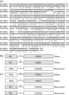
PE_PGRS1 and PE_PGRS33 sequence comparison and constructed chimeras. (a) Protein sequence alignment of PE_PGRS1 and PE_PGRS33 showing the similarities between their PE domains (amino acids 1–100, 62 % homology), their linker regions (italic type, 70 % homology) and PGRS domains (remaining amino acids, 55 % homology). The additional 7 amino acids found in PE_PGRS1 but not PE_PGRS33 are underlined. (b) Schematic of the constructs in which the extra 7 amino acids of PE_PGRS1 were removed and the linker regions of PE_PGRS1 and PE_PGRS33 were swapped, and their localization in transfected RD cells. (c) Schematic of the chimeras in which the PE domains, linker regions and PGRS domains of PE_PGRS1 and PE_PGRS33 were systematically swapped and their localization in transfected RD cells.

References

    1. Abarca-Rojano, E., Rosas-Medina, P., Zamudio-Cortéz, P., Mondragón-Flores, R. & Sánchez-Garcia, F. J Mycobacterium tuberculosis virulence correlates with mitochondrial cytochrome c release in infected macrophages. Scand J Immunol. 2003;58:419–427. - PubMed
    1. Abdallah A. M., Verboom T., Hannes F., Safi M., Strong M., Eisenberg D., Musters R. J., Vandenbroucke-Grauls C. M., Applemelk B. J., other authors A specific secretion system mediates PPE41 transport in pathogenic mycobacteria. Mol Microbiol. 2006;62:667–679. - PubMed
    1. Abdallah A. M., Gey van Pittius N. C., Champion P. A. D., Cox J., Luirink J., Vandenbroucke-Grauls C. M., Applemelk B. J., Bitter W. Type VII secretion – mycobacteria show the way. Nat Rev Microbiol. 2007;5:883–891. - PubMed
    1. Abdallah A. M., Verboom T., Weerdenburg E. M., Gey van Pittius N. C., Mahasha P. W., Jiménez C., Parra M., Cadieux N., Brennan M. J., other authors PPE and PE_PGRS proteins of Mycobacterium marinum are transported via the type VII secretion system ESX-5. Mol Microbiol. 2009;73:329–340. - PubMed
    1. Balaji K. N., Goyal G., Narayana Y., Srinivas M., Chaturvedi R., Mohammad S. Apoptosis triggered by Rv1818c, a PE family gene from Mycobacterium tuberculosis is regulated by mitochondrial intermediates in T cells. Microbes Infect. 2007;9:271–281. - PubMed

Publication types

MeSH terms

LinkOut - more resources