. 2010 Jul 12;11(4):3237.
doi: 10.1120/jacmp.v11i4.3237.

Characterization of Nyquist ghost in EPI-fMRI acquisition sequences implemented on two clinical 1.5 T MR scanner systems: effect of readout bandwidth and echo spacing

Affiliations

Characterization of Nyquist ghost in EPI-fMRI acquisition sequences implemented on two clinical 1.5 T MR scanner systems: effect of readout bandwidth and echo spacing

Marco Giannelli et al. J Appl Clin Med Phys. .

Abstract

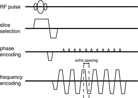
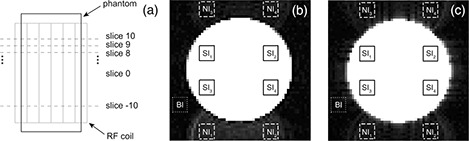
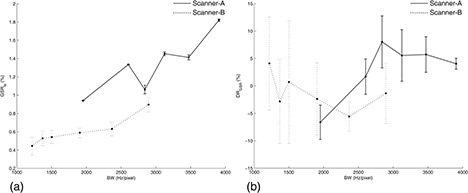
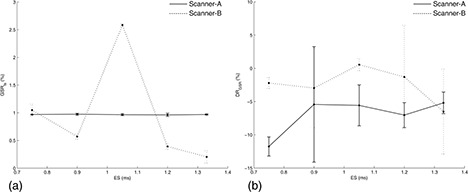
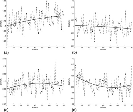
In EPI-fMRI acquisitions, various readout bandwidth (BW) values are used as a function of gradients' characteristics of the MR scanner system. Echo spacing (ES) is another fundamental parameter of EPI-fMRI sequences, but the employed ES value is not usually reported in fMRI studies. Nyquist ghost is a typical EPI artifact that can degrade the overall quality of fMRI time series. In this work, the authors assessed the basic effect of BW and ES for two clinical 1.5 T MR scanner systems (scanner-A, scanner-B) on Nyquist ghost of gradient-echo EPI-fMRI sequences. BW range was: scanner-A, 1953-3906 Hz/pixel; scanner-B, 1220-2894 Hz/pixel. ES range was: scanner-A, scanner-B: 0.75-1.33 ms. The ghost-to-signal ratio of time series acquisition (GSRts) and drift of ghost-to-signal ratio (DRGSR) were measured in a water phantom. For both scanner-A (93% of variation) and scanner-B (102% of variation) the mean GSRts significantly increased with increasing BW. GSRts values of scanner-A did not significantly depended on ES. On the other hand, GSRts values of scanner-B significantly varied with ES, showing a downward trend (81% of variation) with increasing ES. In addition, a GSRts spike point at ES = 1.05 ms indicating a potential resonant effect was revealed. For both scanners, no significant effect of ES on DRGSR was revealed. DRGSR values of scanner-B did not significantly vary with BW, whereas DRGSR values of scanner-A significantly depended on BW showing an upward trend from negative to positive values with increasing BW. GSRts and DRGSR can significantly vary with BW and ES, and the specific pattern of variation may depend on gradients performances, EPI sequence calibrations and functional design of radiofrequency coil. Thus, each MR scanner system should be separately characterized. In general, the employment of low BW values seems to reduce the intensity and temporal variation of Nyquist ghost in EPI-fMRI time series. On the other hand, the use of minimum ES value might not be entirely advantageous when the MR scanner is characterized by gradients with low performances and suboptimal EPI sequence calibration.


Figures

Figure 1
Figure 1
Graphical diagram of gradient‐echo EPI‐fMRI acquisition sequence. The echo spacing represents the interval between successive echoes.
Figure 2
Figure 2
Set up of water phantom acquisitions (a); measurements of GSRts and DRGSR were performed in slice 0 which is the median slice of the acquired phantom volume. Scanner‐A (b): images with BW=1953Hz/pixel and ES=0.9ms. Scanner‐B (c): images with BW=1906Hz/pixel and ES=0.9ms. The ROIs (b, c) used for the measurement of signal intensity within the phantom (SIi,i=1,2,3,4) (solid line), and the ROIs used for the measurement of signal intensity of Nyquist ghost (NIi,i=1,2,3,4) (dashed line) and background (BI) (dotted line), are shown. The ROIs employed for the measurement of NIi(i=1,2,3,4) are shifted by N/2 voxels (64/2) along the phase encoding direction in respect to the corresponding ROIs employed for the measurement of SIi(i=1,2,3,4).
Figure 3
Figure 3
Scanner‐A (solid line): GSRts (a) and DRGSR (b) as a function of BW at fixed ES=0.9ms. Scanner‐B (dotted line): GSRts (a) and DRGSR (b) as a function of BW at fixed ES=0.9ms. Graphs report the mean and standard deviation of the three measurements performed for each BW value.
Figure 4
Figure 4
Scanner‐A (solid line): GSRts (a) and DRGSR (b) as a function of ES at fixed BW=1953Hz/pixel. Scanner‐B (dotted line): GSRts (a) and DRGSR (b) as a function of BW at fixed BW=1502Hz/pixel. Graphs report the mean and standard deviation of the three measurements performed for each ES value.
Figure 5
Figure 5
Temporal variation of ghost‐to‐signal ratio values during EPI‐fMRI acquisitions superimposed with the second order polynomial trend fitted to time series data points: (a) scanner‐A, BW=3125Hz/pixel,ES=0.9ms; (b) scanner‐B, BW=1502Hz/pixel,ES=0.75ms; (c) scanner‐B, BW=1502Hz/pixel,ES=1.05ms; (d) scanner‐A, BW=1953Hz/pixel,ES=0.75ms.

Similar articles

Cited by

  • A longitudinal neuroimaging dataset on multisensory lexical processing in school-aged children.
    Lytle MN, McNorgan C, Booth JR. Lytle MN, et al. Sci Data. 2019 Dec 20;6(1):329. doi: 10.1038/s41597-019-0338-5. Sci Data. 2019. PMID: 31862878 Free PMC article.
  • Radiofrequency artefacts in echoplanar imaging induced by two 1.5 T MR scanners in close proximity.
    Li X, Cui J, Christopasak SP, Kumar A, Peng ZG. Li X, et al. Br J Radiol. 2014 Jun;87(1038):20130773. doi: 10.1259/bjr.20130773. Epub 2014 Apr 9. Br J Radiol. 2014. PMID: 24712321 Free PMC article.
  • Different Neural Activities for Actions and Language within the Shared Brain Regions: Evidence from Action and Verb Generation.
    Wang Z, Xi Q, Zhang H, Song Y, Cao S. Wang Z, et al. Behav Sci (Basel). 2022 Jul 21;12(7):243. doi: 10.3390/bs12070243. Behav Sci (Basel). 2022. PMID: 35877314 Free PMC article.
  • An open resource for transdiagnostic research in pediatric mental health and learning disorders.
    Alexander LM, Escalera J, Ai L, Andreotti C, Febre K, Mangone A, Vega-Potler N, Langer N, Alexander A, Kovacs M, Litke S, O'Hagan B, Andersen J, Bronstein B, Bui A, Bushey M, Butler H, Castagna V, Camacho N, Chan E, Citera D, Clucas J, Cohen S, Dufek S, Eaves M, Fradera B, Gardner J, Grant-Villegas N, Green G, Gregory C, Hart E, Harris S, Horton M, Kahn D, Kabotyanski K, Karmel B, Kelly SP, Kleinman K, Koo B, Kramer E, Lennon E, Lord C, Mantello G, Margolis A, Merikangas KR, Milham J, Minniti G, Neuhaus R, Levine A, Osman Y, Parra LC, Pugh KR, Racanello A, Restrepo A, Saltzman T, Septimus B, Tobe R, Waltz R, Williams A, Yeo A, Castellanos FX, Klein A, Paus T, Leventhal BL, Craddock RC, Koplewicz HS, Milham MP. Alexander LM, et al. Sci Data. 2017 Dec 19;4:170181. doi: 10.1038/sdata.2017.181. Sci Data. 2017. PMID: 29257126 Free PMC article.
  • An Open Resource for Non-human Primate Imaging.
    Milham MP, Ai L, Koo B, Xu T, Amiez C, Balezeau F, Baxter MG, Blezer ELA, Brochier T, Chen A, Croxson PL, Damatac CG, Dehaene S, Everling S, Fair DA, Fleysher L, Freiwald W, Froudist-Walsh S, Griffiths TD, Guedj C, Hadj-Bouziane F, Ben Hamed S, Harel N, Hiba B, Jarraya B, Jung B, Kastner S, Klink PC, Kwok SC, Laland KN, Leopold DA, Lindenfors P, Mars RB, Menon RS, Messinger A, Meunier M, Mok K, Morrison JH, Nacef J, Nagy J, Rios MO, Petkov CI, Pinsk M, Poirier C, Procyk E, Rajimehr R, Reader SM, Roelfsema PR, Rudko DA, Rushworth MFS, Russ BE, Sallet J, Schmid MC, Schwiedrzik CM, Seidlitz J, Sein J, Shmuel A, Sullivan EL, Ungerleider L, Thiele A, Todorov OS, Tsao D, Wang Z, Wilson CRE, Yacoub E, Ye FQ, Zarco W, Zhou YD, Margulies DS, Schroeder CE. Milham MP, et al. Neuron. 2018 Oct 10;100(1):61-74.e2. doi: 10.1016/j.neuron.2018.08.039. Epub 2018 Sep 27. Neuron. 2018. PMID: 30269990 Free PMC article.

References

    1. Ogawa S, Lee TM, Kay AR, Tank DW. Brain magnetic resonance imaging with contrast dependent on blood oxygenation. Proc Natl Acad Sci USA. 1990;87(24):9868–72. - PMC - PubMed
    1. Price RR, Allison J, Massoth RJ, Clarke JD, Drost DJ. Practical aspects of functional MRI (NMR Task Group #8). Med Phys. 2002;29(8):1892–912. - PubMed
    1. Matthews PM, Honey GD, Bullmore ET. Applications of fMRI in translational medicine and clinical practice. Nat Rev Neurosci. 2006;7(9):732–44. - PubMed
    1. Ogawa S, Menon RS, Kim SG, Ugurbil K. On the characteristics of functional magnetic resonance imaging of the brain. Annu Rev Biophys Biomol Struct. 1998;27:447–74. - PubMed
    1. Logothetis NK. What we can do and what we cannot do with fMRI. Nature. 2008;453(7197):869–78. - PubMed

Publication types

MeSH terms