. 2011 Jan 5;30(1):90-103.
doi: 10.1038/emboj.2010.283. Epub 2010 Nov 16.

Smaug assembles an ATP-dependent stable complex repressing nanos mRNA translation at multiple levels

Affiliations

Smaug assembles an ATP-dependent stable complex repressing nanos mRNA translation at multiple levels

Mandy Jeske et al. EMBO J. .

Abstract

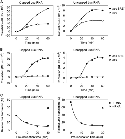
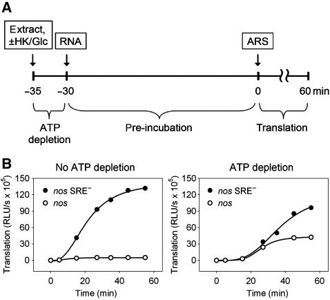
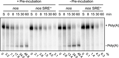
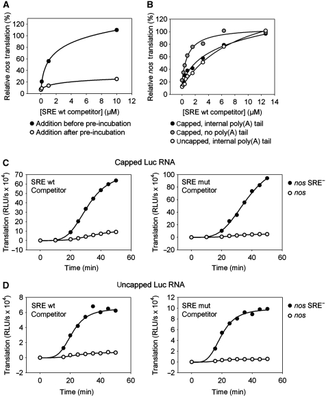
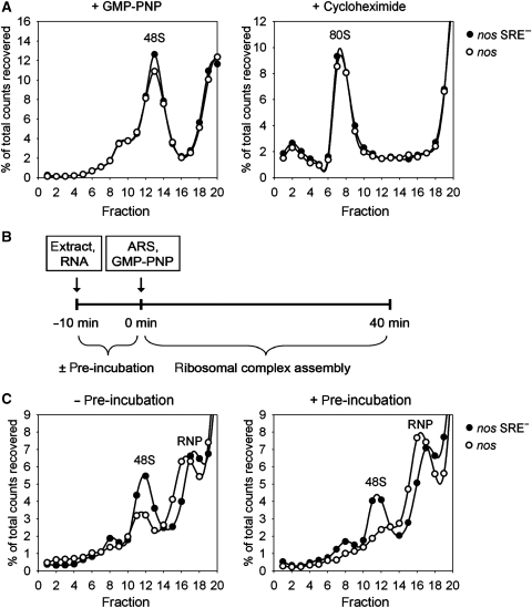
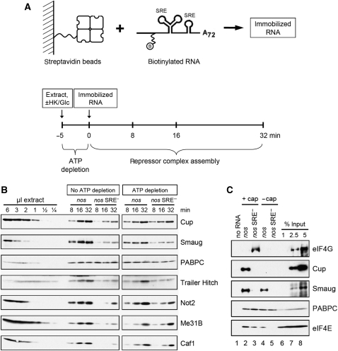
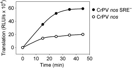
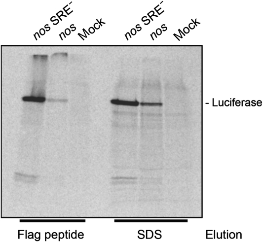
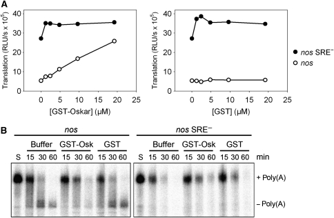
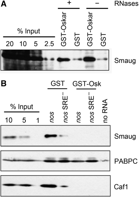
The nanos (nos) mRNA encodes the posterior determinant of the Drosophila embryo. Translation of the RNA is repressed throughout most of the embryo by the protein Smaug binding to Smaug recognition elements (SREs) in the 3' UTR. Translation is locally activated at the posterior pole by Oskar. This paper reports that the SREs govern the time- and ATP-dependent assembly of an exceedingly stable repressed ribonucleoprotein particle (RNP) in embryo extract. Repression can be virtually complete. Smaug and its co-repressor Cup as well as Trailer hitch and the DEAD box protein Me31B are part of the repressed RNP. The initiation factor eIF4G is specifically displaced, and 48S pre-initiation complex formation is inhibited. However, later steps in translation initiation are also sensitive to SRE-dependent inhibition. These data confirm several previously untested predictions of a current model for Cup-dependent repression but also suggest that the Cup model by itself is insufficient to explain translational repression of the nos RNA. In the embryo extract, recombinant Oskar relieves translational repression and deadenylation by preventing Smaug's binding to the SREs.


Conflict of interest statement

The authors declare that they have no conflict of interest.

Figures

Figure 1
Figure 1
Efficient repression depends on the slow assembly of a repressor complex. Capped (left panels) or uncapped (right panels) luciferase RNAs containing an internal poly(A) tract were incubated in embryo extract under translation conditions. At the time points indicated, the translation yield was determined by a luciferase assay. (A) Luciferase RNAs containing the nos 3′ UTR fragment (circles) or the fragment with point mutations in the SRE sequences (nos SRE, filled circles) were mixed with extract, and translation was started immediately. (B) The same experiment as in (A) was carried out, but luciferase RNAs were pre-incubated with extract for 30 min before translation was started by the addition of the ATP-regenerating system (see Materials and methods) at t=0. (C) Luciferase RNAs containing the nos or the nos SRE 3′ UTR fragment were pre-incubated with extract for the times indicated before translation was started. Luciferase activity obtained from the SRE RNA after 1 h of translation was set to 100%. The circle indicates the relative translation level obtained from a reaction where luciferase RNAs were added after the pre-incubation step.
Figure 2
Figure 2
Translation repression is ATP dependent. (A) Scheme of the assay: embryo extract was incubated with hexokinase and glucose for 5 min for ATP depletion. Capped luciferase RNAs containing an internal poly(A) tract were then added and pre-incubated for 30 min. Subsequently, translation was started by addition of the ATP-regenerating system (ARS). The control reaction was performed similarly, but the extract was initially incubated for 5 min without addition of hexokinase and glucose. (B) Luciferase RNAs containing the nos (circles) or the nos SRE (filled circles) 3′ UTR fragment were incubated as described in (A). For a better comparison, the ordinates of the two graphs are drawn in the same scale. The time scale refers to the start of translation.
Figure 3
Figure 3
SRE-dependent deadenylation is not accelerated by a pre-incubation step. Polyadenylated, uncapped, and 32P-labelled RNA substrates containing the nos or the nos SRE 3′ UTR fragment were subjected to a 25-min pre-incubation with Drosophila embryo extract, then deadenylation was started by addition of the ATP-regenerating system (0 min; left half of the panel). After the time points indicated, RNA was isolated and separated on a denaturing polyacrylamide gel. The control reaction (right half of the panel) was performed in the same way but the pre-incubation step was omitted. S, unreacted substrate RNA. The percentage of fully deadenylated product (Jeske et al, 2006) is shown below each lane.
Figure 4
Figure 4
The assembled repressor complex is stable. (A) Capped luciferase RNAs containing an internal poly(A) tract and the nos or the nos SRE 3′ UTR fragment were pre-incubated for 25 min and subsequently translated for 60 min. Increasing amounts of a short-competitor SRE RNA were added to the reaction either before (filled circles) or after (circles) the pre-incubation step. The percentage of translation of the SRE-containing RNA relative to the mutant RNA is plotted against the competitor concentration. (B) Luciferase RNAs containing an internal poly(A) tract with the indicated 5′ and 3′ end modifications were mixed with increasing amounts of the SRE competitor. Subsequently, the RNA mixture was pre-incubated for 25 min with embryo extract followed by a 60-min translation reaction. The percentage of translation of the SRE-containing RNA relative to the mutant RNA is plotted against the competitor concentration. (C) Capped luciferase RNAs containing an internal poly(A) tract and the nos (circles) or the nos SRE (filled circles) 3′ UTR fragment were pre-incubated for 25 min with embryo extract to allow repressor complex formation on nos RNA. After pre-incubation, 12 μM of wild-type (left) or mutant (right) SRE competitor was added to the mixture together with the ATP-regenerating system (t=0 min), and a time course of translation was measured. (D) The same experiment as described in (C) was performed using uncapped luciferase RNAs.
Figure 5
Figure 5
Repressed nos RNA does not form 48S complexes. (A) 32P-cap-labelled RNAs containing an internal poly(A) tract and the indicated 3′ UTR were incubated with rabbit reticulocyte lysate in the presence of cycloheximide (left panel) or GMP-PNP (right panel) under translation conditions. Resulting ribosomal complexes were resolved by centrifugation in 5–25% linear sucrose gradients (25% sucrose on left). After fractionation from the bottom to the top of the gradient, the radioactivity was monitored, expressed as percentage of total counts recovered and plotted against the fraction number. (B) Scheme of ribosomal complex assembly reaction using Drosophila embryo extract. 32P-cap-labelled RNAs containing an internal poly(A) tract were incubated under translation conditions in the presence of GMP-PNP either with or without a preceding pre-incubation step for 10 min. ARS, ATP-regenerating system. (C) Substrate RNAs with the indicated 3′ UTR were allowed to assemble ribosomal complexes as described in (B) and analysed as described in (A).
Figure 6
Figure 6
RNA pull-down assays reveal components of the repressed RNP: exclusion of eIF4G. (A) Scheme of the RNA pull-down assay. Biotinylated and 32P-labelled RNA substrates containing an external poly(A) tail were immobilized on streptavidin beads (upper panel). Embryo extract was incubated for 5 min in the presence of hexokinase and glucose for ATP depletion or without these reagents as a control. Immobilized RNA was added and incubated with the extract for the times indicated (lower panel). The beads were washed and bound proteins eluted by boiling in SDS sample buffer. Proteins were separated on an SDS–polyacrylamide gel and analysed by western blotting. (B) Proteins were detected in an RNA pull-down assay as described in (A) using capped nos and nos SRE RNA substrates. In all, 36 μl of embryo extract were used for each time point of the pull down. For comparison, various amounts of extract were analysed in the same western blot (shown in the first six lanes). (C) eIF4G is excluded from the repressed RNP. An RNA pull-down assay was carried out as described in (A) except that no ATP depletion was carried out. Capped and uncapped substrate RNAs carrying the nos or the nos SRE sequences were compared as indicated. Immobilized RNAs were incubated with embryo extract for 30 min. Beads without immobilized RNA were used as a background control.
Figure 7
Figure 7
CrPV IRES-dependent translation is repressed in an SRE-dependent manner. Uncapped luciferase reporter RNAs containing the CrPV-IRES, the indicated 3′ UTR and internal poly(A) tails were pre-incubated for 30 min with embryo extract under the conditions described in Materials and methods. Subsequently, translation was started by the addition of an ATP-regenerating system, and luciferase activity was measured at the time points indicated.
Figure 8
Figure 8
SRE-dependent repression has no effect on nascent peptide chains. Capped luciferase reporter RNAs with an N-terminal flag tag and an internal poly(A) tract were translated as in Figure 1B. 35S-Methionine was added at 2 μCi/reaction. Translation reactions were stopped with 5 μl 50 mM EDTA and 1 mg/ml RNase A, incubated for 20 min at 30°C and mixed with 85 μl IP buffer (150 mM NaCl, 50 mM Tris–HCl pH 8.0, 0.5% Triton X-100, 1 mg/ml yeast total RNA). The mixture was transferred to 20 μl anti-FLAG agarose (Sigma) and incubated for 40 min at 8°C with shaking. The agarose was washed five times with 200 μl IP buffer. Proteins were sequentially eluted with FLAG peptide (1 mg/ml in IP buffer) and SDS-loading buffer and separated on a 10% SDS–PAGE. The picture shows the autoradiogram of the gel. ‘Mock' is a translation reaction without added reporter RNA.
Figure 9
Figure 9
Recombinant Oskar prevents both SRE-dependent translation repression and deadenylation. (A) Translation reaction mixtures lacking RNA and the ATP-regenerating system were incubated with increasing amounts of recombinant GST-Oskar (left panel) or GST (right panel) for 15 min. Capped luciferase RNAs containing an internal poly(A) tract and with the indicated 3′ UTR fragments were then added, and the incubation continued for another 25 min before translation was started by the addition of the ATP-regenerating system and allowed to proceed for 60 min. Luciferase activity is plotted against concentration of the protein indicated. When no protein was added (0 μM), protein buffer was used instead. (B) Deadenylation reaction mixtures were incubated for 15 min with GST-Oskar, GST, or protein buffer in the absence of RNA and an ATP-regenerating system. Deadenylation was started by addition of polyadenylated, 32P-labelled substrate RNAs with the 3′ UTR fragments indicated and buffer containing the ATP-regenerating system. At the time points indicated, RNA was isolated and separated on a denaturing polyacrylamide gel. S, unreacted substrate RNA.
Figure 10
Figure 10
Recombinant Oskar associates with Smaug and prevents its binding to the SRE. (A) Oskar binds to Smaug independently of RNA. Embryo extract was incubated with GST-Oskar or GST in the presence or absence of RNases (see Materials and methods). After capture of the proteins on glutathione beads and washing, bound proteins were eluted by boiling in SDS sample buffer. Samples were separated on an SDS–polyacrylamide gel and analysed by western blotting. Different amounts of the input extract were loaded for comparison. (B) Oskar prevents binding of Smaug to SREs. An RNA pull-down assay was carried out as described in Figure 6 (A) using capped RNAs with the 3′ UTR fragments indicated. Embryo extract was incubated with GST-Oskar or GST for 15 min, immobilized RNAs were added and incubated with the mixture for another 30 min. Beads without RNA were used as background control. Bound proteins were analysed by western blotting with antibodies against Smaug, CAF1, and PABPC as indicated. Different amounts of the input extract were loaded for comparison.

Similar articles

Cited by

References

    1. Aviv T, Lin Z, Ben-Ari G, Smibert CA, Sicheri F (2006) Sequence-specific recognition of RNA hairpins by the SAM domain of Vts1p. Nat Struct Mol Biol 13: 168–176 - PubMed
    1. Aviv T, Lin Z, Lau S, Rendl LM, Sicheri F, Smibert CA (2003) The RNA binding SAM domain of Smaug defines a new family of post-transcriptional regulators. Nat Struct Biol 10: 614–621 - PubMed
    1. Barbee SA, Estes PS, Cziko A-M, Hillebrand J, Luedeman RA, Coller JM, Johnson N, Howlett IC, Geng C, Ueda R, Brand AH, Newbury SF, Wilhelm JE, Levine RB, Nakamura A, Parker R, Ramaswami M (2006) Staufen- and FMRP-containing neuronal RNPs are structurally and functionally related to somatic P bodies. Neuron 52: 997–1009 - PMC - PubMed
    1. Bashirullah A, Halsell SR, Cooperstock RL, Kloc M, Karaiskakis A, Fisher WW, Fu W, Hamilton JK, Etkin LD, Lipshitz HD (1999) Joint action of two RNA degradation pathways controls the timing of maternal transcript elimination at the midblastula transition in Drosophila melanogaster. EMBO J 18: 2610–2620 - PMC - PubMed
    1. Bergsten SE, Gavis ER (1999) Role for mRNA localization in translational activation but not spatial restriction of nanos RNA. Development 126: 659–669 - PubMed

Publication types

MeSH terms