. 2011 Mar;19(3):450-60.
doi: 10.1038/mt.2010.249. Epub 2010 Nov 16.

Minicircle DNA-based gene therapy coupled with immune modulation permits long-term expression of α-L-iduronidase in mice with mucopolysaccharidosis type I

Affiliations

Minicircle DNA-based gene therapy coupled with immune modulation permits long-term expression of α-L-iduronidase in mice with mucopolysaccharidosis type I

Mark J Osborn et al. Mol Ther. 2011 Mar.

Abstract

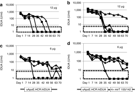
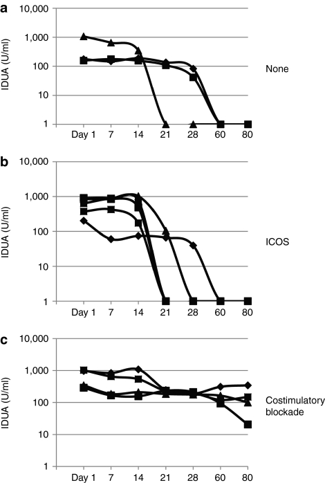
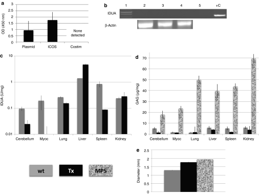
Mucopolysaccharidosis type I (MPS I) is a lysosomal storage disease characterized by mutations to the α-L-iduronidase (IDUA) gene resulting in inactivation of the IDUA enzyme. The loss of IDUA protein results in the progressive accumulation of glycosaminoglycans within the lysosomes resulting in severe, multi-organ system pathology. Gene replacement strategies have relied on the use of viral or nonviral gene delivery systems. Drawbacks to these include laborious production procedures, poor efficacy due to plasmid-borne gene silencing, and the risk of insertional mutagenesis. This report demonstrates the efficacy of a nonintegrating, minicircle (MC) DNA vector that is resistant to epigenetic gene silencing in vivo. To achieve sustained expression of the immunogenic IDUA protein we investigated the use of a tissue-specific promoter in conjunction with microRNA target sequences. The inclusion of microRNA target sequences resulted in a slight improvement in long-term expression compared to their absence. However, immune modulation by costimulatory blockade was required and permitted for IDUA expression in MPS I mice that resulted in the biochemical correction of pathology in all of the organs analyzed. MC gene delivery combined with costimulatory pathway blockade maximizes safety, efficacy, and sustained gene expression and is a new approach in the treatment of lysosomal storage disease.


Figures

Figure 1
Figure 1
Plasmid DNA expression in vivo is rapidly lost. Cytomegalovirus (CMV)-regulated transgene expression in immune-deficient mice. (a) CMV promoter driven expression cassette with the human α--idurondase gene and the BGH polyadenylation signal (BpA). Plasmid components include an ampicillin and neomycin resistance gene, the pUC origin of replication and SV40 regulatory elements. (b) α--Iduronidase (IDUA) enzymatic activity was assessed in the plasma of immune-deficient NOD-SCIDγc mice at which point the first group (black bar) was killed and liver genomic DNA harvested. A 24 hour and 7 day time point was collected for plasma IDUA on a second group (gray bars) followed by elective sacrifice and isolation of genomic DNA. Plasmid copy number. (c) A Taqman copy number assay was used by diluting known amounts of the CMV IDUA plasmid to generate a standard curve plotting cycle threshold (Ct) (x axis) against log plasmid copy number (y axis) with y = −0.2816x + 11.905; R2 = 0.99968. (d) Plasmid copy numbers in animals 1 or 7 days postinjection. (e) Plasma IDUA levels of animals treated with the original 13.2 µg dose compared to animals treated with one half of that amount (6.6 µg) corresponding to the average loss of DNA copies in d. Black, hatched box indicates the normal level of IDUA present in a wild-type mouse. N = 3.
Figure 2
Figure 2
IDUA-based minicircle cassette mechanism, purification, function, and maintenance as an episome in vivo. (a) Minicircle (MC) expression cassette and generation. The expression cassette, comprised of an enhancer promoter containing the human apolipoprotein E hepatic locus control region and the human α-1-antitrypsin promoter (sApoE.HCR.hAATp) driving human α--iduronidase (hIDUA) production with the bovine growth hormone polyadenylation signal (bpA), is flanked by the φ-C31 attachment B (attB) and attachment P (attP) sites. The phiC31 recombinase gene is present in two copies (BAD C31x2) and it and the I-Sce I Ig gene are under the control of the arabinose inducible BAD promoter. The I-Sce I homing endonuclease site is denoted by the cross. Amp, ampicillin resistance gene; pUC, pUC plasmid origin of replication; araC, araC repressor. (i) The full-length prerecombination plasmid is approximately 12 kb. The addition of arabinose and incubation at 32 °C for 2 hours results in the activation of the phiC31 genes and the generation of two products: (ii) the bacterial backbone (iii) the recombined MC. Supplemental arabinose is added followed by an increase of the culture temperature to 37 °C for an additional 2 hours resulting in the activation of the I-Sce I gene causing linearization and degradation of the backbone elements. (b) Agarose gel electrophoresis of the plasmid in its prerecombination, FL state labeled “pre”. The bands shown are the linearized and supercoiled species. The postrecombination MC species is shown at right. The differing plasmid species in the postrecombination MC are the: (i) ~12 kb linearized full length, (ii) bacterial backbone elements, and (iii) the ~3 kb MC, with the MC representing the major (~86%) product. (c) Treatment of mice with the unrecombined, full length (FL) plasmid or the MC. NOD-SCIDγc mice (n = 4 for FL and n = 3 for MC) received equimolar amounts of either DNA species and plasma α--iduronidase (IDUA) levels were measured at the specified time points. Maintenance of the MC as an episomal species. Liver genomic DNA from the MC treated animals in 2c was isolated 50 days postinjection and analyzed by inverse PCR. The diagram above the agarose gel (d) show the primer design whereby human IDUA specific primers are pointing away from one another resulting in amplification of a DNA species that is circular. The PCR products were resolved on an agarose gel (d). The same liver total genomic DNA was digested with BglII, an enzyme that cuts the MC or FL once, and hybridized with a human IDUA specific probe. Lane 1 of d and e are molecular weight (mw) standards, lanes 2–4 are the individual animals treated with MC from 2c, lane 5 is a positive control containing plasmid DNA spiked into liver genomic DNA and lane 6 is a the negative control containing untreated mouse liver DNA. Black, hatched box indicates the normal level of IDUA present in a wild-type mouse.
Figure 3
Figure 3
MC gene transfer in vivo into immune competent C57Bl/6 mice in the absence and presence of immunomodulation. C57Bl/6 mice were injected with minicircle (MC) in the absence of immune depletion or with targeted depletion of a specific arm of the immune system followed by MC injection and plasma α--iduronidase (IDUA) activity. (a) No immune depletion. (b) NK-cell depletion with PK136 (anti-NK1.1) antibody. (c) CD4 depletion with GK1.5 antibody. (d) CD8 depletion with 2.43 antibody. (e) CD4 and CD8 cell depletion with GK1.5 and 2.43. Arrow indicates the point when immunomodulation was withdrawn. Black, hatched box is the endogenous level of IDUA in wild-type mice. N = 4 each group.
Figure 4
Figure 4
micro RNA regulated IDUA MC gene transfer in vivo. Recombined minicircle (MC) with the IDUA 3′ untranslated region (UTR) containing four copies of each of the miRNA target site (mirT) for (a) mir155 (b) mir-142-3p or (c) 155/142 miRNA was injected into C57Bl/6 mice (n = 4/group) and plasma α--iduronidase (IDUA) levels were assessed weekly. Black, hatched box is the endogenous level of IDUA in wild-type mice.
Figure 5
Figure 5
Plasmid dosage effect on transgene expression. Recombined minicircle (MC) without or with the IDUA 3′ untranslated region (UTR) containing mirT for 155/142 miRNA was injected into C57Bl/6 mice (n = 4/group) at dosages of 12 µg (a and b) or 6 µg (c and d) with plasma α--iduronidase (IDUA) levels assessed weekly. Black, hatched box is the endogenous level of IDUA in wild-type mice.
Figure 6
Figure 6
MPS I MC gene transfer in vivo with immunomodulation. MPS I mice (~6 months of age) were injected with the minicircle (MC)-IDUA mirT142/155 expression cassette and plasma IDUA activity was assessed. (a) IDUA activity in MPS I mice (n = 3) in the absence of immune depletion. (b) Plasma IDUA in mucopolysaccharidosis type I (MPS I) mice (n = 6) that received inducible costimulator (ICOS) (7E.1769.67) antibody. (c) Plasma IDUA (n = 4) in mice that received costimulatory blockade with anti-B7.1 (16-10A1), anti-B7.2 (GL-1), and anti-CD40 ligand (MR-1).
Figure 7
Figure 7
Humoral and innate immune response in MPS mice, organ IDUA enzyme and glycosaminoglycan levels and assessment of skeletal pathology. (a) Humoral immune response. Sera from the treatment groups was assessed for the presence of anti-IDUA antibodies by incubating a 1:50 dilution with recombinant human IDUA that was adsorbed to a microtiter plate. IgG antibody was then detected by adding a goat anti-mouse horse radish peroxidase conjugated antibody and monitoring the color development at Abs 450. (b) Innate immune response. Liver genomic DNA from animals that received minicircle (MC) α--iduronidase (IDUA) with inducible costimulator (ICOS) treatment or costimulatory blockade were assessed for the presence of IDUA vector genomes by PCR. Lane 1 is the molecular weight standard, lanes 2–4 are liver genomic DNA from ICOS treated mice, lane 5 is the no template control and lane 6 is liver genomic DNA from a mouse that showed detectable levels of plasma IDUA. Lower bands are PCR products for β-actin that served as a quality control. (c) Tissue IDUA and GAG Levels. Homogenates from wild-type (n = 3), untreated MPS I −/− mice (n = 4) and mucopolysaccharidosis type I (MPS I) mice receiving IDUA MC in conjunction with costimulatory blockade (n = 4) were analyzed for IDUA enzyme activity (c) and (d) biochemically for glycosaminoglycan (GAG) content. The means and SEM are shown. Levels are normalized to protein and for all tissues in untreated MPS mice versus treated MPS mice the P value was P < 0.01 (Student's t-test). (e) Skeletal pathology. The femora of age matched, sex matched wild-type, MPS, and treated animals (~9 months of age) were measured at the midshaft point. For MPS control mice versus treated MPS mice the P value was P = 0.0003 (Student's t-test). The small amount of substrate converted in the IDUA enzyme assay by untreated MPS mice was subtracted from the remaining groups. The OD value from untreated MPS mice for the antibody assay was subtracted from all groups. ‘Wt', ‘Tx', and ‘MPS' denotes wild type, treated, or untreated MPS control mice, respectively.

Similar articles

Cited by

References

    1. Hacein-Bey-Abina S, Garrigue A, Wang GP, Soulier J, Lim A, Morillon E, et al. Insertional oncogenesis in 4 patients after retrovirus-mediated gene therapy of SCID-X1. J Clin Invest. 2008;118:3132–3142. - PMC - PubMed
    1. Chen ZY, Riu E, He CY, Xu H., and, Kay MA. Silencing of episomal transgene expression in liver by plasmid bacterial backbone DNA is independent of CpG methylation. Mol Ther. 2008;16:548–556. - PubMed
    1. Riu E, Grimm D, Huang Z., and, Kay MA. Increased maintenance and persistence of transgenes by excision of expression cassettes from plasmid sequences in vivo. Hum Gene Ther. 2005;16:558–570. - PubMed
    1. Thomas CE, Ehrhardt A., and, Kay MA. Progress and problems with the use of viral vectors for gene therapy. Nat Rev Genet. 2003;4:346–358. - PubMed
    1. Yew NS., and, Cheng SH. Reducing the immunostimulatory activity of CpG-containing plasmid DNA vectors for non-viral gene therapy. Expert Opin Drug Deliv. 2004;1:115–125. - PubMed

Publication types

MeSH terms