. 2010 Nov 3;5(11):e15428.
doi: 10.1371/journal.pone.0015428.

Expression and membrane topology of Anopheles gambiae odorant receptors in lepidopteran insect cells

Affiliations

Expression and membrane topology of Anopheles gambiae odorant receptors in lepidopteran insect cells

Panagiota Tsitoura et al. PLoS One. .

Abstract

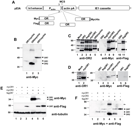
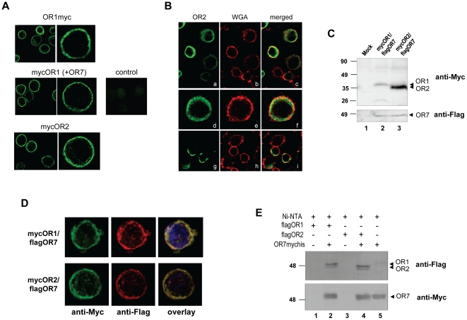
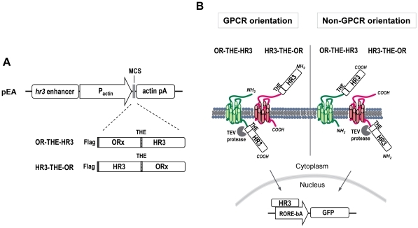
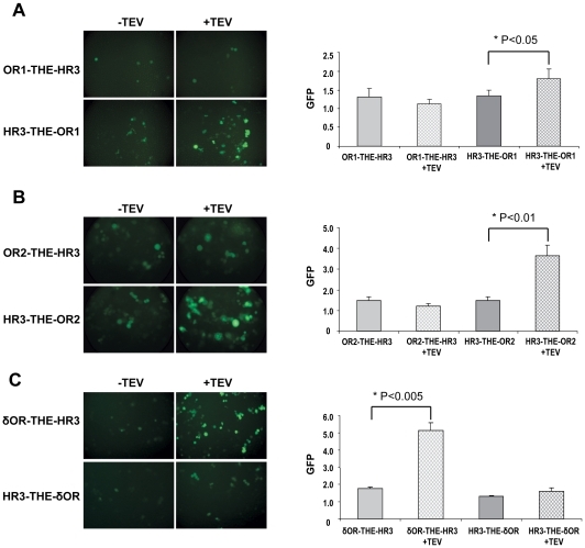
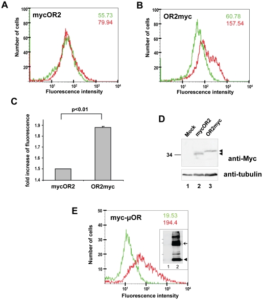
A lepidopteran insect cell-based expression system has been employed to express three Anopheles gambiae odorant receptors (ORs), OR1 and OR2, which respond to components of human sweat, and OR7, the ortholog of Drosophila's OR83b, the heteromerization partner of all functional ORs in that system. With the aid of epitope tagging and specific antibodies, efficient expression of all ORs was demonstrated and intrinsic properties of the proteins were revealed. Moreover, analysis of the orientation of OR1 and OR2 on the cellular plasma membrane through the use of a novel 'topology screen' assay and FACS analysis demonstrates that, as was recently reported for the ORs in Drosophila melanogaster, mosquito ORs also have a topology different than their mammalian counterparts with their N-terminal ends located in the cytoplasm and their C-terminal ends facing outside the cell. These results set the stage for the production of mosquito ORs in quantities that should permit their detailed biochemical and structural characterization and the exploration of their functional properties.


Conflict of interest statement

Competing Interests: The authors have declared that no competing interests exist.

Figures

Figure 1
Figure 1. Expression of A. gambiae OR1, OR2 and OR7 in insect cells.
(A) Schematic representation of the basic backbone vector (pEIA) used for the heterologous expression of various forms (tagged and untagged) of ORs in lepidopteran cells. hr3 enhancer, baculoviral (BmNPV) homologous region 3 enhancer sequence; pActin, Bombyx mori A3 cytoplasmic actin promoter; MCS, multiple cloning site; actin pA, 3′untranslated region of B. mori actin gene containing polyadenylation signals; IE1 cassette, baculoviral (BmNPV) DNA fragment containing the ie-1 transactivator gene under the control of its native viral promoter; OR; A. gambiae odorant receptor ORF; Myc, Flag and MycHis, epitope tags. (B) Detection of heterologous expression of C-terminally MycHis-tagged ORs in transfected Hi5 cells using Myc monoclonal antibody. (C) Detailed western blot analysis of OR2. Hi5 cells were transfected with plasmids expressing different versions of OR2, and lysates were analyzed using a specific polyclonal antibody against OR2 (left panel, lanes 1–5) or monoclonal antibodies against the Myc (middle panel, lanes 6–9) or the Flag epitope (right panel, lanes 10–11). Arrowheads and arrows indicate major bands corresponding to monomers and putative dimers, respectively. (D) Detailed western blot analysis of OR1. Hi5 cells were transfected with plasmids expressing different versions of OR1, and lysates were analyzed using monoclonal antibodies against the Myc (middle panel, lanes 1–4) or the Flag epitope (right panel, lanes 5–6). In the left panel immunoreactivity of the specific polyclonal antibody against OR1 is shown, with lysates from cells expressing mycOR1 after treatment with the proteasome inhibitor MG132. Molecular weight markers are shown on the left. (E) and (F) Effect of coexpression of OR7 on the expression levels of OR1 and OR2. Hi5 cells were transfected with constant amounts (45% of total DNA) of Myc-tagged OR1 or OR2, along with equal amounts of Flag-tagged OR7 or empty vector (pEIA), and pEIA-GFP (10% of total DNA, for evaluation of the efficiency of transfection). Whole cell lysates (E) and membrane fractions (F) were analysed by SDS-PAGE and western blot. Detection of OR1, OR2 and OR7 was done using the anti-Myc and anti-Flag antibodies either consecutively (in E) or simultaneously (in F). To control for loading, the whole lysate fractions were also probed with an anti-tubulin antibody.
Figure 2
Figure 2. Co-localization of odorant receptors expressed in lepidopteran insect cells.
(A) Expression constructs for N-terminally (mycOR1, mycOR2) or C-terminally tagged receptors (OR1myc) were transfected in Bm5 cells (in the absence/presence of OR7) and the localization of the expressed ORs was detected using anti-Myc antibody. Control indicates transfection with empty expression vector. (B) Co-localization of OR2 with the plasma membrane marker wheat germ agglutinin. Cells expressing OR2myc were double stained with WGA-Texas Red-X conjugate (b, e, f) and with anti-myc antibody (a, d, g) in the presence or absence of saponin (a-f and g-i, respectively). (C) Detection of ORs in the membrane fraction of stable cell lines coexpressing mycOR1 (lane 2) or mycOR2 (lane 3) along with flagOR7. Immunoblotting was performed with anti-Myc and anti-Flag antibodies (upper and lower panels, respectively). Membranes from Bm5 untransfected cells were used as a negative control (lane 1). (D) Co-localization of OR1 or OR2 with OR7. Bm5 cells were co-transfected with expression plasmids for N-terminally tagged mycOR1 or mycOR2 together with N-terminally tagged flagOR7 expression vector. Tagged ORs were detected with anti-Myc/anti-mouse fluorescein-labelled IgG and anti-Flag/anti-rabbit Alexa fluor-labelled IgG as indicated and counter-stained with DAPI. (E) Pull-down assays showing heteromerization between OR1 and OR7 or OR2 and OR7. Extracts containing C-terminally Myc-His-tagged OR7 were incubated with Ni2+-NTA beads and bound protein complexes were analyzed by Western blot by anti-Flag antibody (upper panel) for the presence of N-terminally Flag-tagged OR1 and OR2 or by anti-Myc antibody (lower panel) to detect OR7mychis.
Figure 3
Figure 3. The “topology screen” assay.
(A) Expression constructs. hr3 enhancer, baculoviral (BmNPV) homologous region 3 enhancer sequence; pActin, Bombyx mori A3 cytoplasmic actin promoter; MCS, multiple cloning site; actin pA, 3′untranslated region of B. mori actin gene containing polyadenylation signals; Flag, epitope tag; OR; Odorant or opioid receptor ORF; THE, Tobacco etch virus protease recognition site; HR3, Bombyx mori hormone receptor 3. (B) Hypothetical model illustrating possible location of HR3 in both fusion constructs, with respect to the OR orientation (GPCR or not) in the membrane. RORE-bA, response element for retinoic acid receptor-related orphan receptor/basal actin promoter.
Figure 4
Figure 4. Topology assays for the mosquito OR1 and OR2.
Chimeric receptor proteins fused at either their N- or the C- terminus to the TEV cleavage sequence (THE) and the HR3 transcription factor are co-expressed in Hi5 cells with a GFP reporter construct together with or without TEV protease. For both OR1 (A) and OR2 (B) N-terminal fusions (HR3-THE-OR1 and HR3-THE-OR2), expression of the TEV protease resulted in increase of fluorescence of the cells. No significant increase of fluorescence was detected when the C-terminal fusions of ORs were used (OR1-THE-HR3 and OR2-THE-HR3). Similar constructs of the human opioid receptor δ (δOR) that was used as control (C) give opposite results with an increased fluorescence for the C-terminal fusion (δOR-THE-HR3). For each chimeric receptor protein, both representative images (left) and quantitative results (right, with values representing the mean ± S.E.M. of four experiments) from the fluorometric analysis are shown.
Figure 5
Figure 5. Flow cytometric analysis of expression of Myc-tagged OR2 on the surface of Bm5 cells.
(A) N- or (B) C-terminally Myc-tagged OR2 were stably expressed in Bm5 cells in the presence of flagOR7 and analyzed by FACS for the extracellular localization of the Myc tag. For each panel, the green tracing and number represent fluorescence values obtained from the staining of the cells with only the FITC labelled secondary antibodies, while the red tracing and number represent values obtained from cells incubated with both the primary anti-Myc and the FITC labelled secondary antibodies. Increased fluorescence intensity (2.59-fold over the background value) was observed for the C-terminally Myc-tagged receptor in comparison to the receptor that was Myc-tagged at the N-terminal end (1.43-fold over the background value). (C) Values (increases over background) indicated in this panel represent the mean ± S.E.M. of three independent experiments. (D) Western blot analyses of whole cell lysates from the stable cell lines used for FACS analysis and control, mock-transformed cells, probed with anti-Myc (upper) and anti- tubulin (lower) antibodies. (E) FACS analysis of cells expressing the N-terminally Myc-tagged µ-opioid receptor used as a positive control for the extracellular localization of the Myc tag. Green and red tracing/numbers are as in panels A and B. Inset shows the detection of µOR in these cells by western blotting with the anti-myc antibody. The arrowhead and the arrow point to major bands detected (putative monomer and dimer, respectively), while the positions of 50, 90 and 118-kDa molecular mass markers are indicated at left.

Similar articles

Cited by

References

    1. Rajagopal S, Rajagopal K, Lefkowitz RJ. Teaching old receptors new tricks: biasing seven-transmembrane receptors. Nat Rev Drug Discov. 2010;9:373–386. - PMC - PubMed
    1. Bargmann CI. Comparative chemosensation from receptors to ecology. Nature. 2006;444:295–301. - PubMed
    1. Lundin C, Kall L, Kreher SA, Kapp K, Sonnhammer EL, et al. Membrane topology of the Drosophila OR83b odorant receptor. FEBS Lett. 2007;581:5601–5604. - PMC - PubMed
    1. Wistrand M, Kall L, Sonnhammer EL. A general model of G protein-coupled receptor sequences and its application to detect remote homologs. Protein Sci. 2006;15:509–521. - PMC - PubMed
    1. Benton R, Sachse S, Michnick SW, Vosshall LB. Atypical membrane topology and heteromeric function of Drosophila odorant receptors in vivo. PLoS Biol. 2006;4:e20. - PMC - PubMed

Publication types

MeSH terms